Deep intrabony defects treated using Straumann® Emdogain® - Dr. M. Stefanini
-
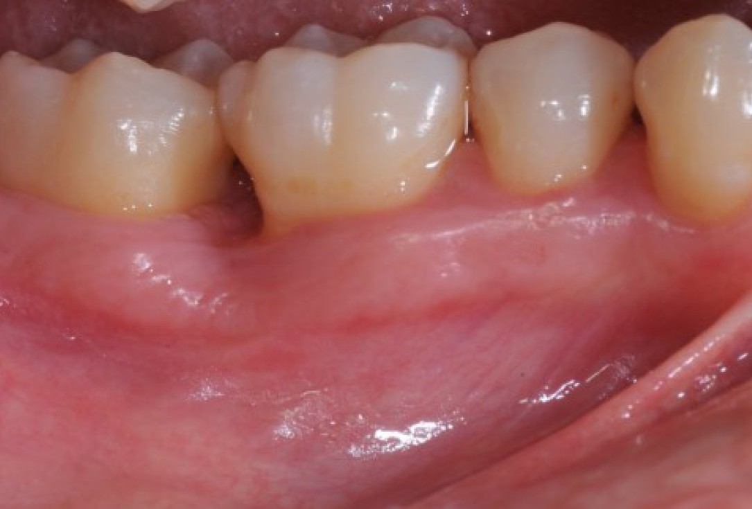
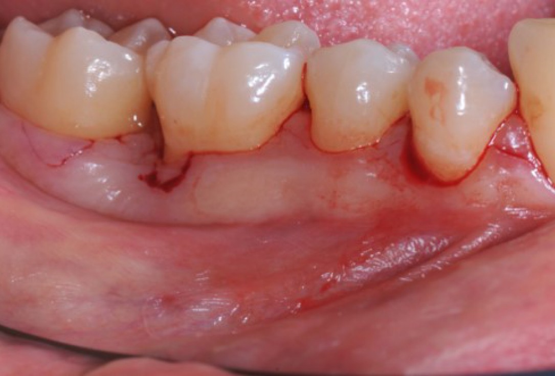
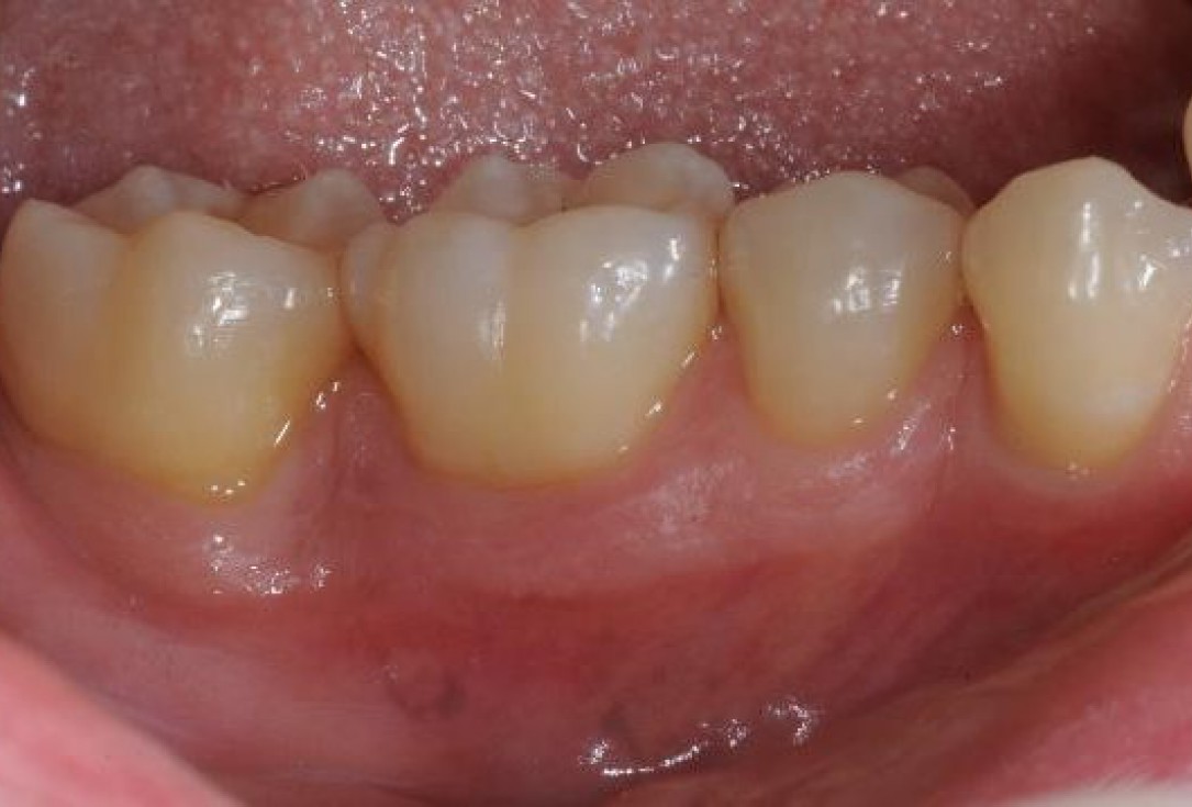
01/22 - Pre-surgical clinical situation, buccal view.Deep intrabony defects treated using Straumann® Emdogain® - Dr. M. Stefanini
-
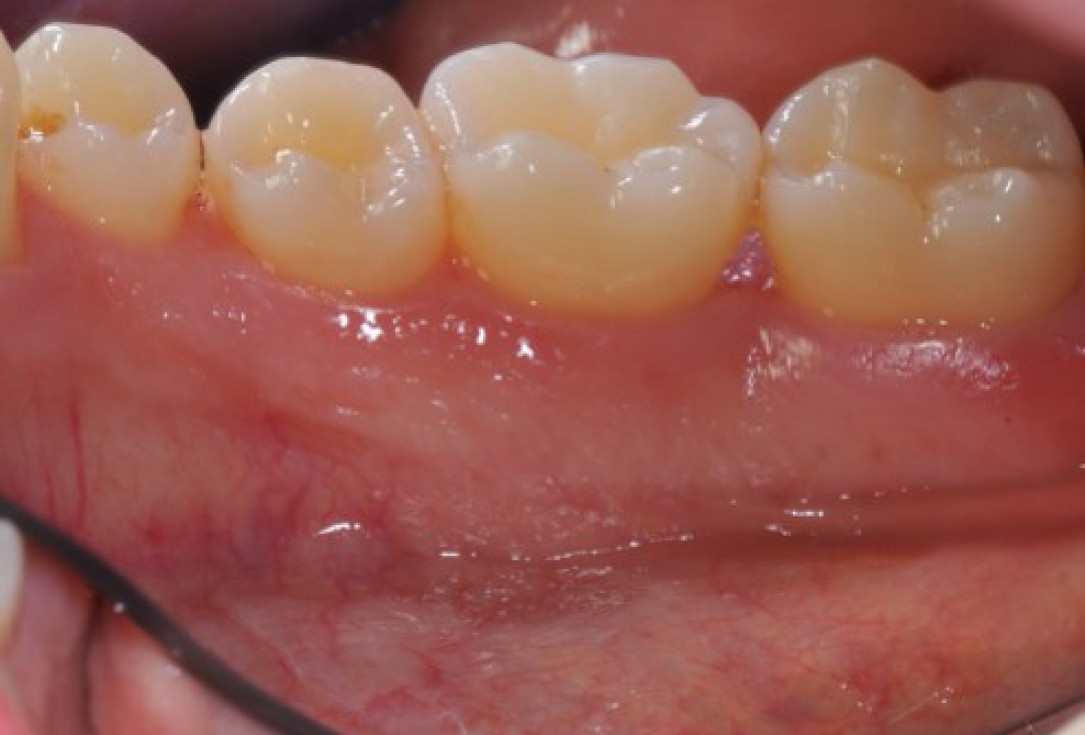
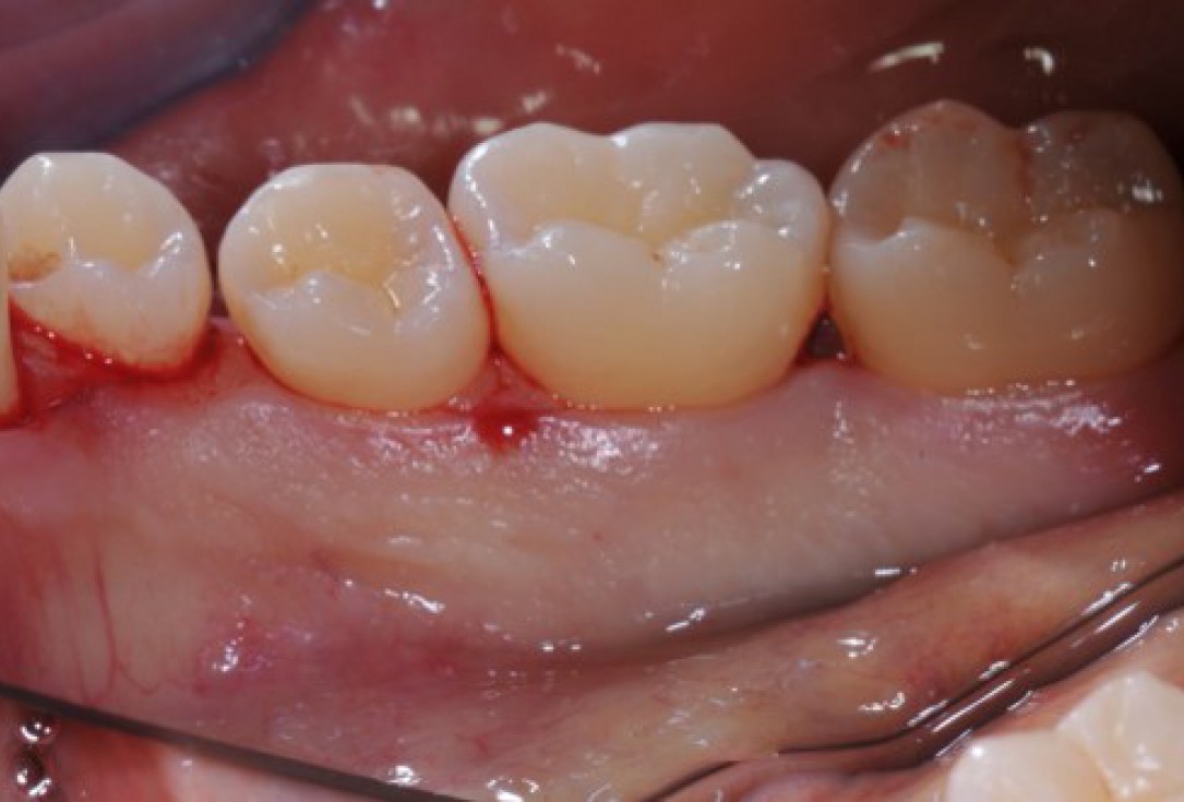
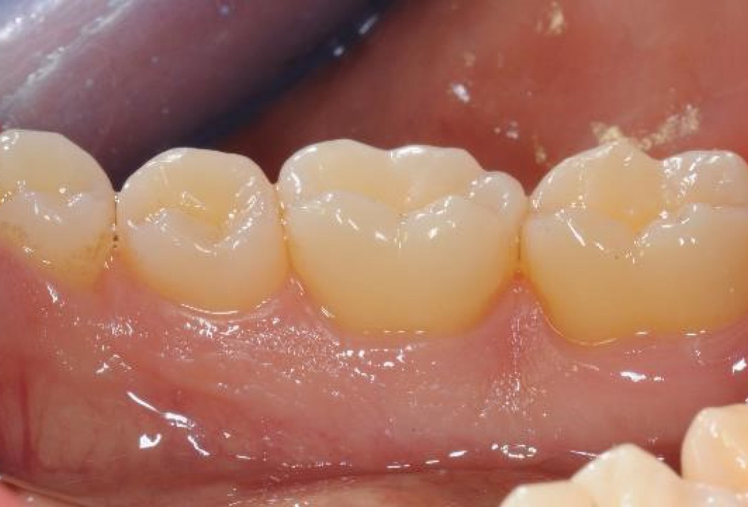
02/22 - Pre-surgical clinical situation, lingual view.Deep intrabony defects treated using Straumann® Emdogain® - Dr. M. Stefanini
-
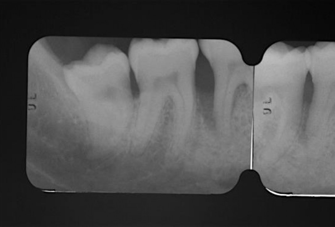
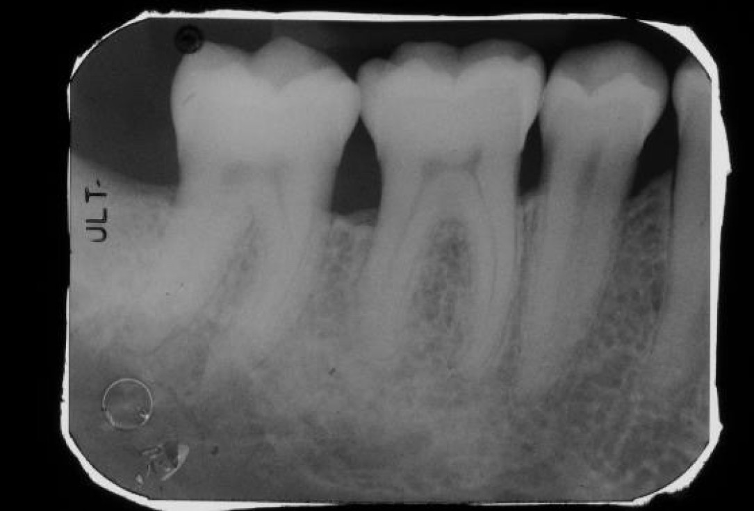
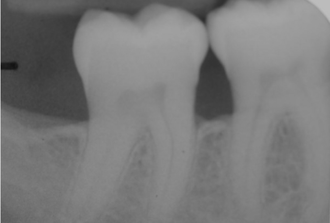
03/22 - Pre-operative radiograph. Deep intrabony defect visible on the distal aspect of tooth 46.Deep intrabony defects treated using Straumann® Emdogain® - Dr. M. Stefanini
-
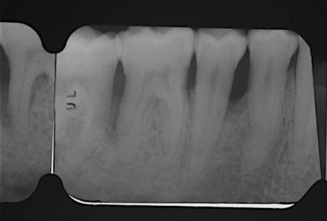
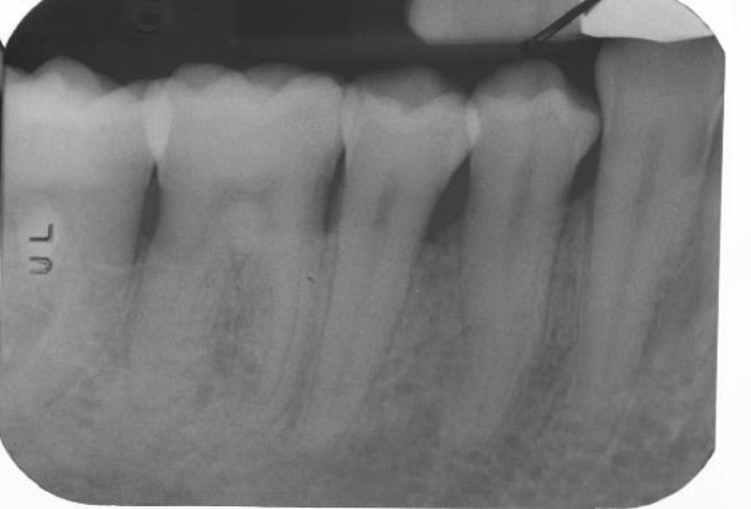
04/22 - Pre-operative radiograph. Deep intrabony defect visible mesially to tooth 45.Deep intrabony defects treated using Straumann® Emdogain® - Dr. M. Stefanini
-
05/22 - Access flap (simplified papilla preservation technique between #44 and #45 and amplified papilla preservation technique between #47 and# 46). Buccal view.Deep intrabony defects treated using Straumann® Emdogain® - Dr. M. Stefanini
-
06/22 - Access flap (simplified papilla preservation technique between #44 and #45 and amplified papilla preservation technique between #47 and# 46). Lingual view.Deep intrabony defects treated using Straumann® Emdogain® - Dr. M. Stefanini
-
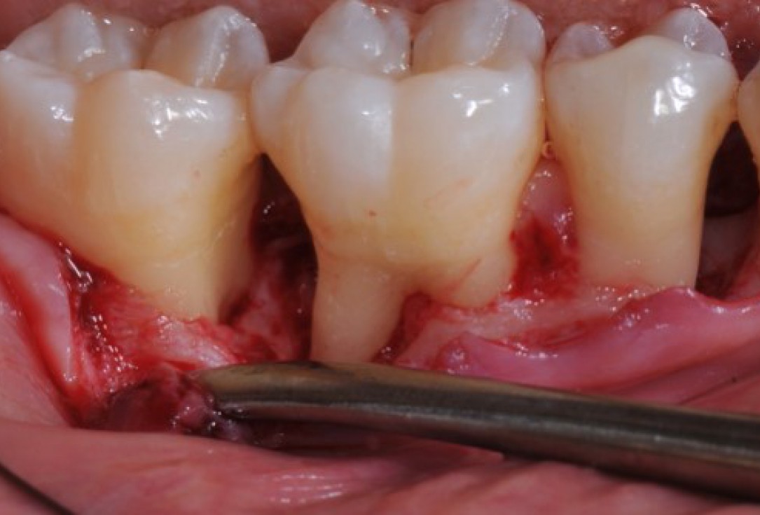
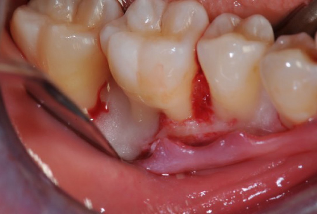
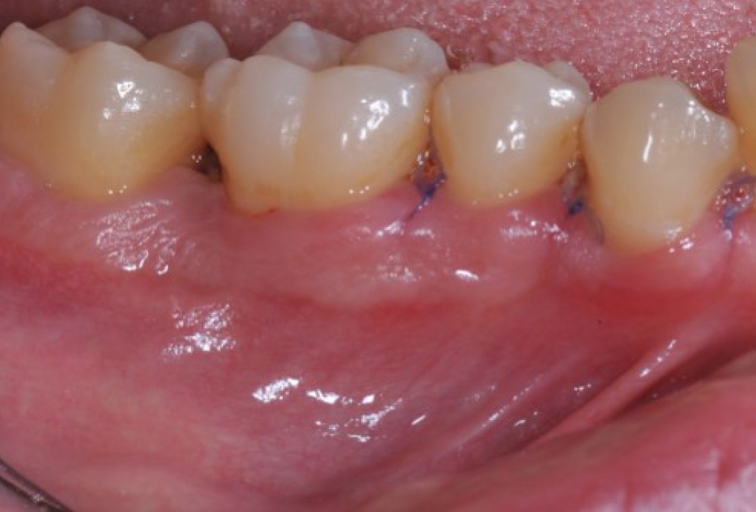
07/22 - Intra-operative view reveals deep non-contained intrabony defects distally to tooth 46 (PPD 10 mm) and on the mesial aspect of tooth 45 (PPD 6 mm). Buccal view.Deep intrabony defects treated using Straumann® Emdogain® - Dr. M. Stefanini
-
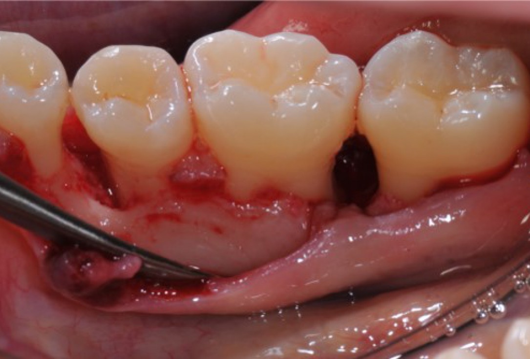
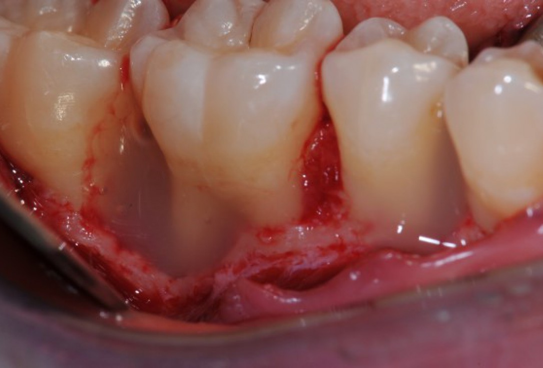
08/22 - Intra-operative view reveals deep non-contained intrabony defects distally to tooth 46 (PPD 10 mm) and on the mesial aspect of tooth 45 (PPD 6 mm). Lingual view.Deep intrabony defects treated using Straumann® Emdogain® - Dr. M. Stefanini
-
09/22 - Application of Straumann® PrefGel® all over the exposed root surfaces.Deep intrabony defects treated using Straumann® Emdogain® - Dr. M. Stefanini
-
10/22 - After thorough rinsing, application of Straumann® Emdogain® onto the exposed root surface.Deep intrabony defects treated using Straumann® Emdogain® - Dr. M. Stefanini
-
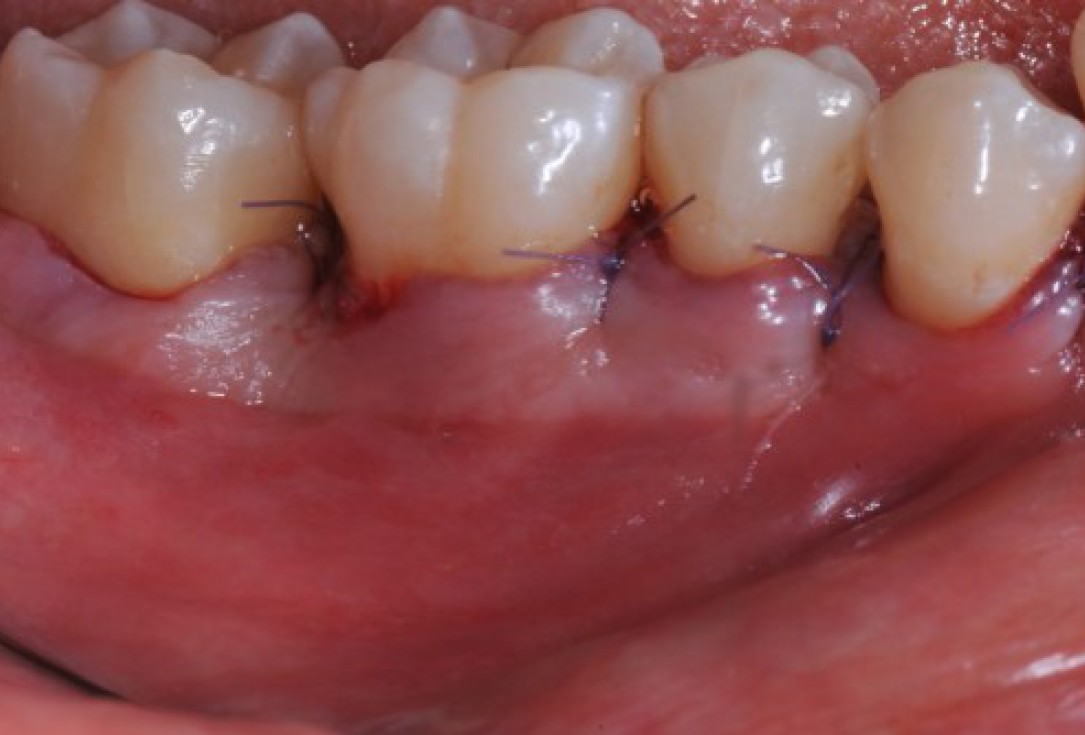
11/22 - Coronal advancement of the flap and suturing to achieve primary wound closure. Buccal view.Deep intrabony defects treated using Straumann® Emdogain® - Dr. M. Stefanini
-
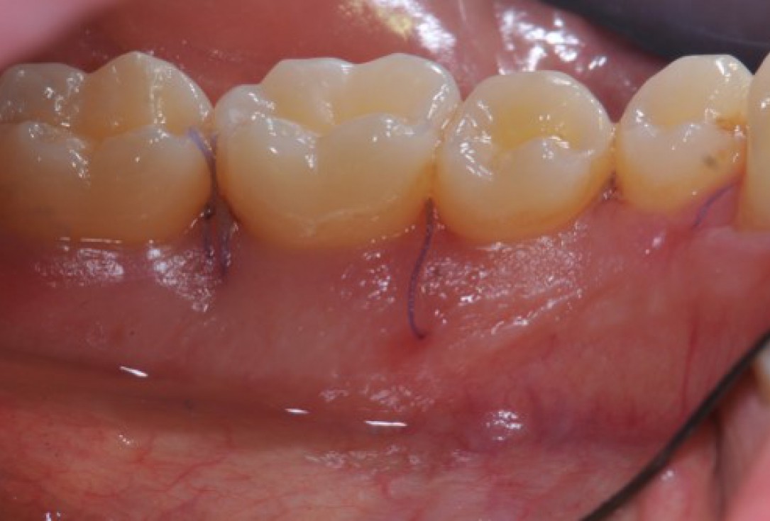
12/22 - Coronal advancement of the flap and suturing to achieve primary wound closure. Lingual view.Deep intrabony defects treated using Straumann® Emdogain® - Dr. M. Stefanini
-
13/22 - Removal of the sutures 14 days post-operative. Buccal view.Deep intrabony defects treated using Straumann® Emdogain® - Dr. M. Stefanini
-
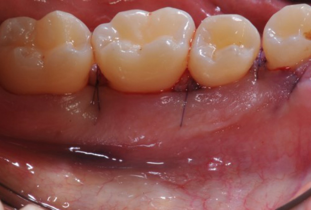
14/22 - Removal of the sutures 14 days post-operative. Lingual view.Deep intrabony defects treated using Straumann® Emdogain® - Dr. M. Stefanini
-
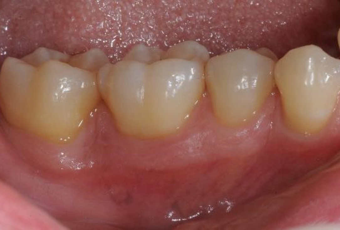
15/22 - Clinical situation 12 months post-operative. Buccal view.Deep intrabony defects treated using Straumann® Emdogain® - Dr. M. Stefanini
-
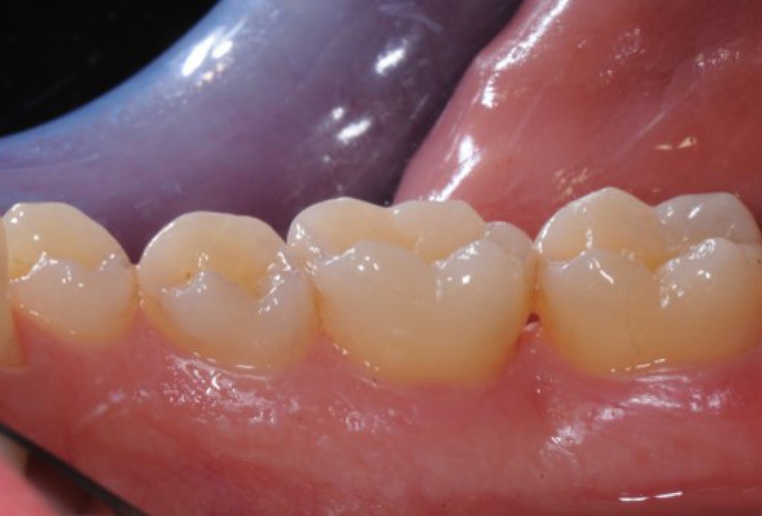
16/22 - Clinical situation 12 months post-operative. Lingual view.Deep intrabony defects treated using Straumann® Emdogain® - Dr. M. Stefanini
-
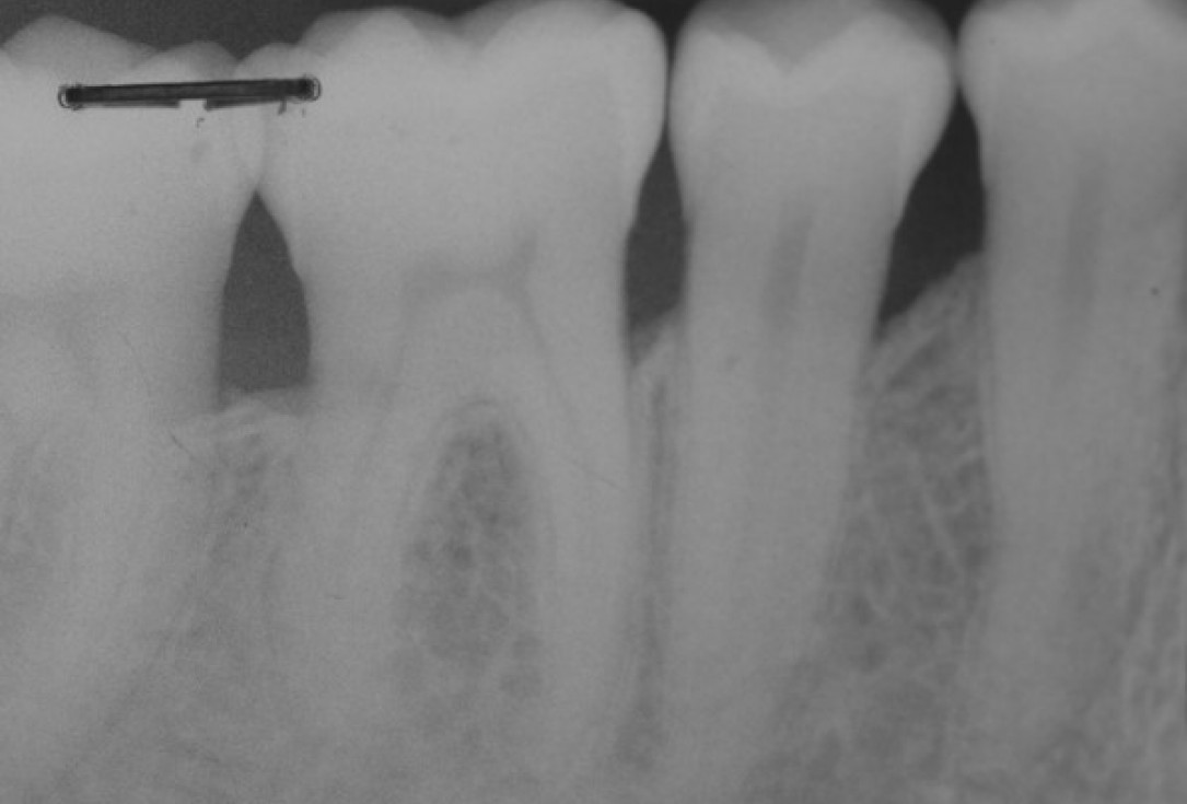
17/22 - Radiographic follow up 12 months post-operative. The radiograph demonstrates a complete defect fill and a stable result.Deep intrabony defects treated using Straumann® Emdogain® - Dr. M. Stefanini
-
18/22 - Radiographic follow up 12 months post-operative. The radiograph demonstrates a complete defect fill and a stable result.Deep intrabony defects treated using Straumann® Emdogain® - Dr. M. Stefanini
-
19/22 - Long term follow up: clinical situation 6 years post-operative. Buccal view.Deep intrabony defects treated using Straumann® Emdogain® - Dr. M. Stefanini
-
20/22 - Long term follow up: clinical situation 6 years post-operative. Lingual view.Deep intrabony defects treated using Straumann® Emdogain® - Dr. M. Stefanini
-
21/22 - Long term follow up: radiographic situation 6 years post-operative.Deep intrabony defects treated using Straumann® Emdogain® - Dr. M. Stefanini
-
22/22 - Long term follow up: radiographic situation 6 years post-operative.Deep intrabony defects treated using Straumann® Emdogain® - Dr. M. Stefanini

Alveolar socket before soft and hard tissue augmentation

Pre-operative OPG shows deep vertical intrabony defects on the distal aspects of teeth 13 and 14.

Radiographic view before periodontal regenerative therapy with Straumann® Emdogain®. A deep intrabony defect appeared mesially and distally on the left mandibular first premolar. Pre-surgical probing measured 8 mm. The defect morphology presented as well-contained.

Baseline clincial situation and pre-surgical probing.

Pre-operative radiograph. Intrabony defect on the mesial aspect of tooth 14.

Pre-operative clinical situation. Gingival recessions at teeth 11 and 21.

Pre-surgical situation. Multiple adjacent gingival recessions at teeth 12, 13 and 14.

Pre-operative clinical view. Multiple adjacent gingival recessions.

Initial clinical situation

Pre-operative X-ray. Hopless tooth 21.

Pre-operative clinical situation.

Pre-operative probing pocket depth (PPD) at the distal aspect of tooth 11 was 7 mm.

Pre-surgical clinical situation. Deep gingival recessions at both upper canine.

Pre-operative clinical situation. Multiple adjacent gingival recessions.

Pre-operative clinical situation. Shallow multiple adjacent gingival recessions in the first quadrant.

Initial situation: bone loss due to lack of physical load of bridge retained region 11

Pre-operative radiographic view. Intrabony defect on the distal aspect of the lateral incisor.

Situation after tooth removal.

Pre-operative clinical situation.

Pre-operative radiographic view.

Baseline clinical situation. Recession depth of 6 mm at tooth 31.

Initial situation: 40 year old female patient with extensive scar tissue after several surgeries restored with a Rochette bridge
