Vertical bone augmentation and broadening of attached gingiva using cerabone®, permamem® and mucoderm® - Dr. R. Naimoli
-
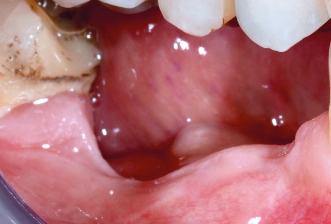
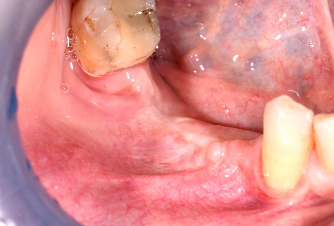
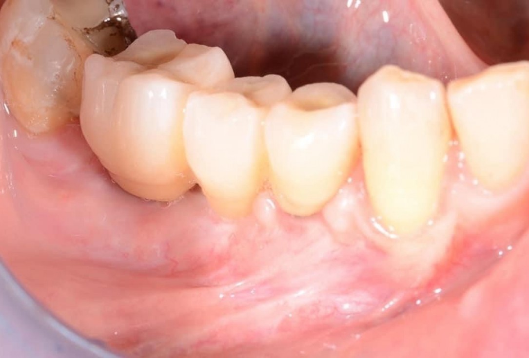
01/29 - Initial clinical situation with pronounced vertical and horizontal bone defectVertical bone augmentation and broadening of attached gingiva using cerabone®, permamem® and mucoderm® - Dr. R. Naimoli
-
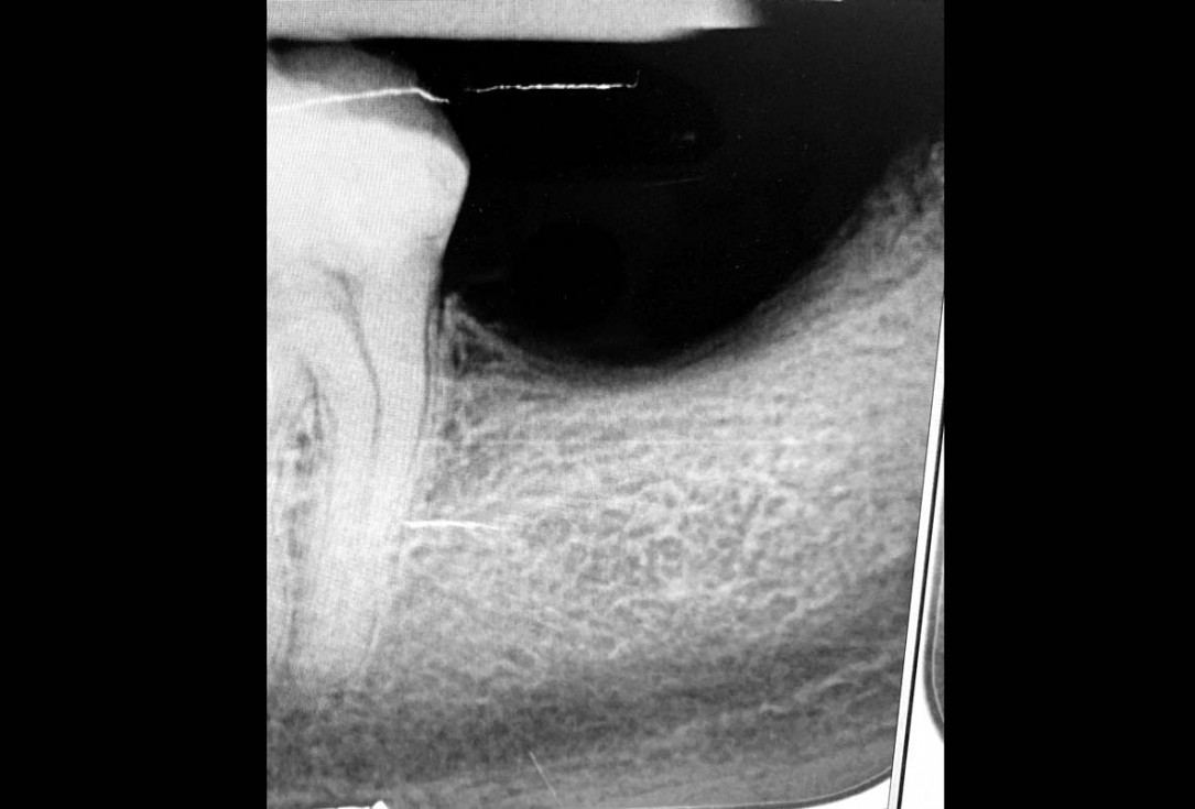
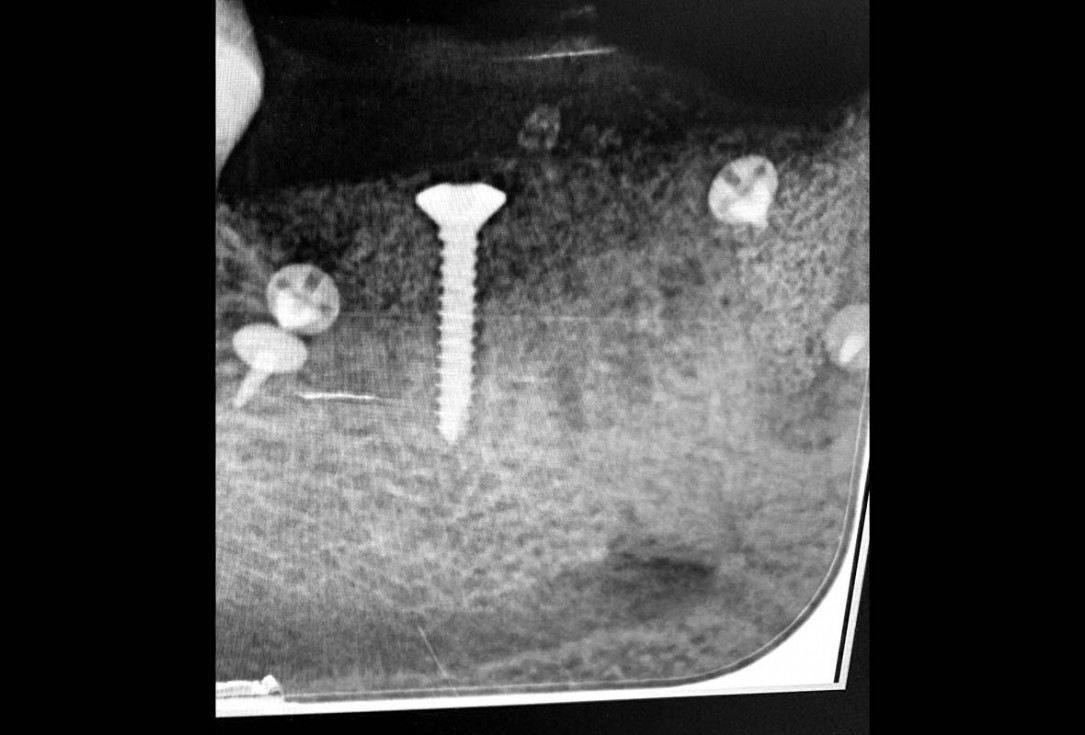
02/29 - Based on the intraoral x-ray a vertical bone defect of about 4 mm extending from 4.6 to 4.4 was determinedVertical bone augmentation and broadening of attached gingiva using cerabone®, permamem® and mucoderm® - Dr. R. Naimoli
-
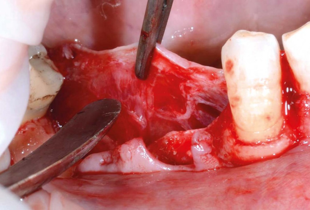
03/29 - Preparation of a full-thickness flapVertical bone augmentation and broadening of attached gingiva using cerabone®, permamem® and mucoderm® - Dr. R. Naimoli
-
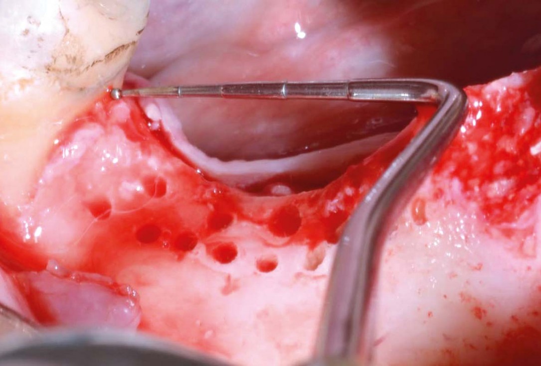
04/29 - Clinical view after exposure of the defect, to promote healing the local bone was perforatedVertical bone augmentation and broadening of attached gingiva using cerabone®, permamem® and mucoderm® - Dr. R. Naimoli
-
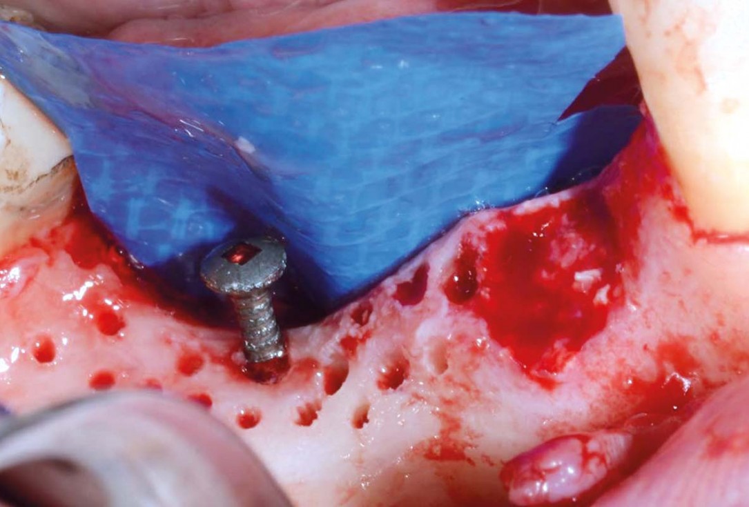
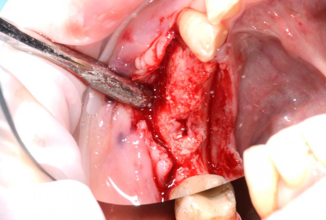
05/29 - Fixation of an osteosynthesis screw at the lowest point of the defect, lingual positioning of permamem®Vertical bone augmentation and broadening of attached gingiva using cerabone®, permamem® and mucoderm® - Dr. R. Naimoli
-
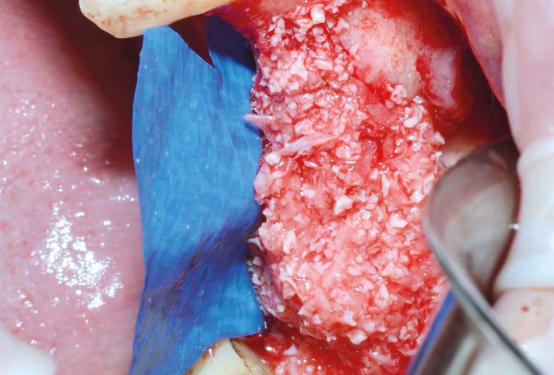
06/29 - Augmentation with cerabone® and autologous bone (ratio of 1:1)Vertical bone augmentation and broadening of attached gingiva using cerabone®, permamem® and mucoderm® - Dr. R. Naimoli
-
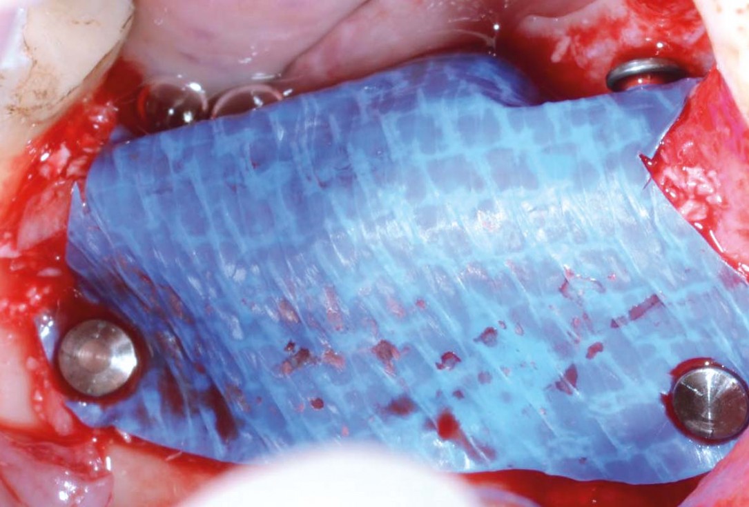
07/29 - Covering of the augmented area and buccal fixation of permamem® with titanium pinsVertical bone augmentation and broadening of attached gingiva using cerabone®, permamem® and mucoderm® - Dr. R. Naimoli
-
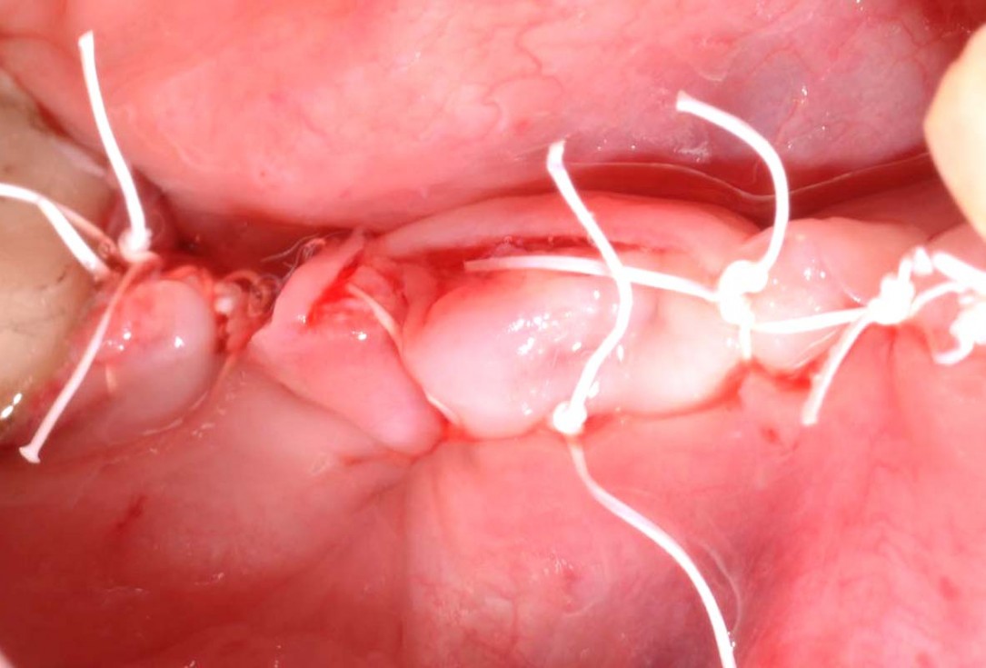
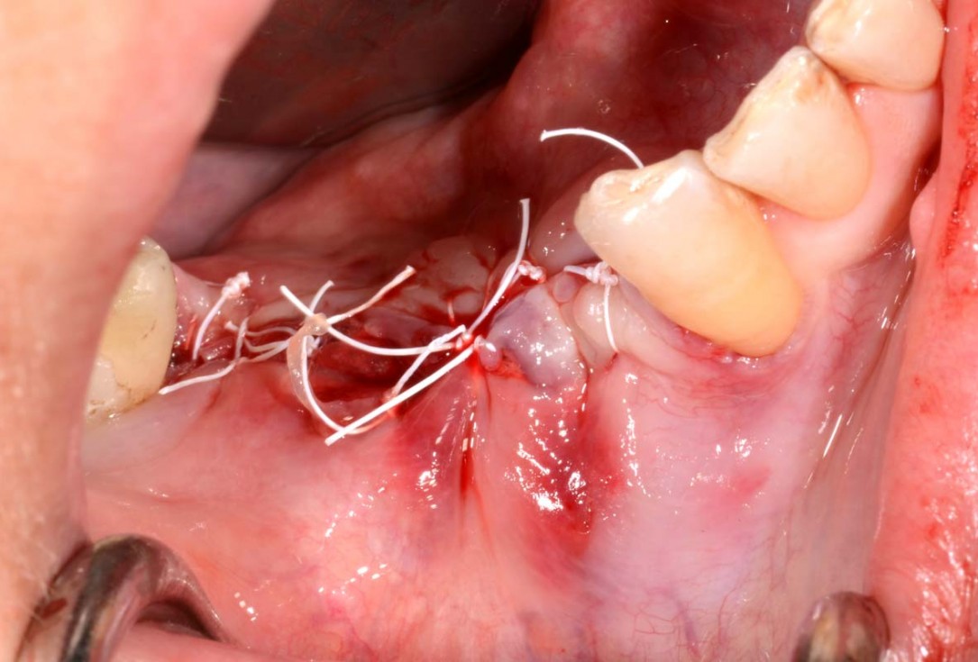
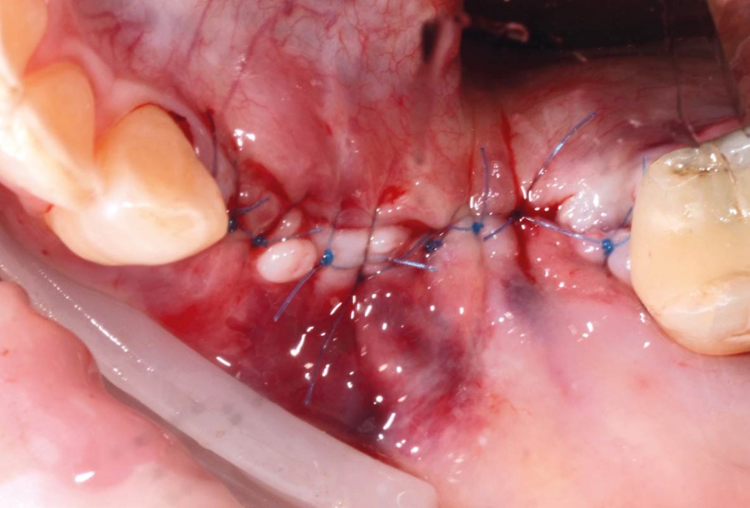
08/29 - Defect closure with non-absorbable PTFE suturesVertical bone augmentation and broadening of attached gingiva using cerabone®, permamem® and mucoderm® - Dr. R. Naimoli
-
09/29 - Post-operative x-ray controlVertical bone augmentation and broadening of attached gingiva using cerabone®, permamem® and mucoderm® - Dr. R. Naimoli
-
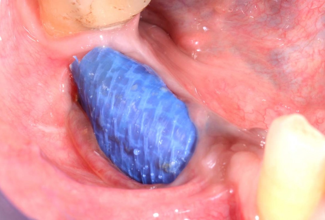
10/29 - Dehiscence of permamem® 3 months post-operatively. No signs of infection, the patient did not report painVertical bone augmentation and broadening of attached gingiva using cerabone®, permamem® and mucoderm® - Dr. R. Naimoli
-
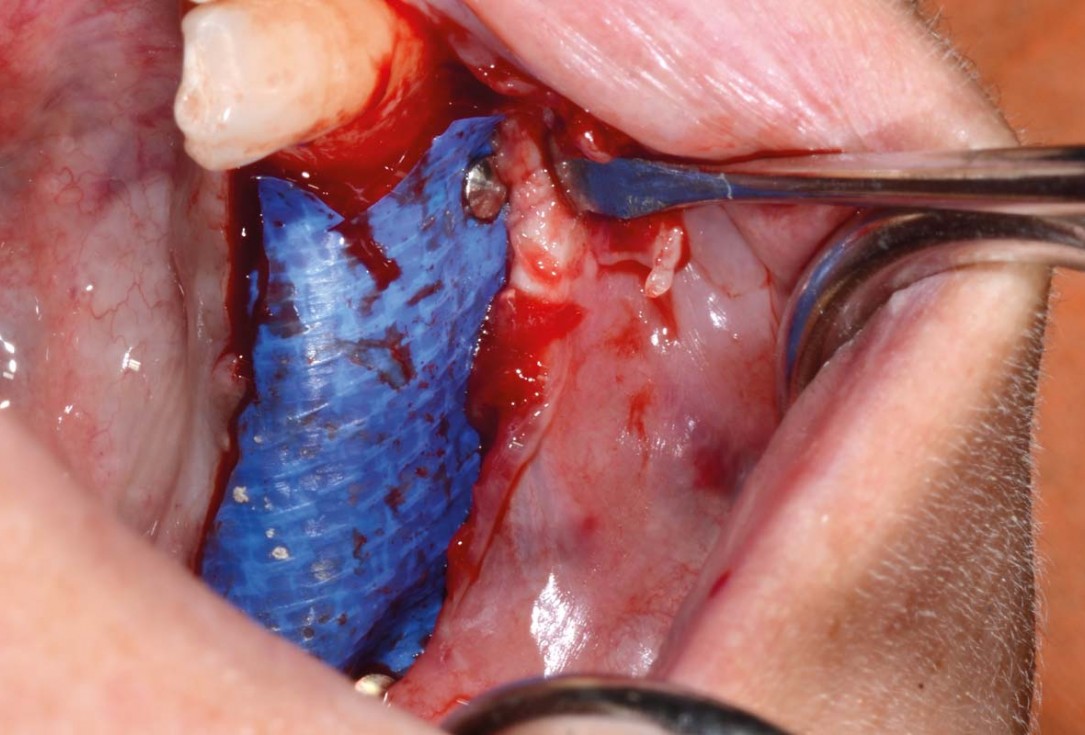
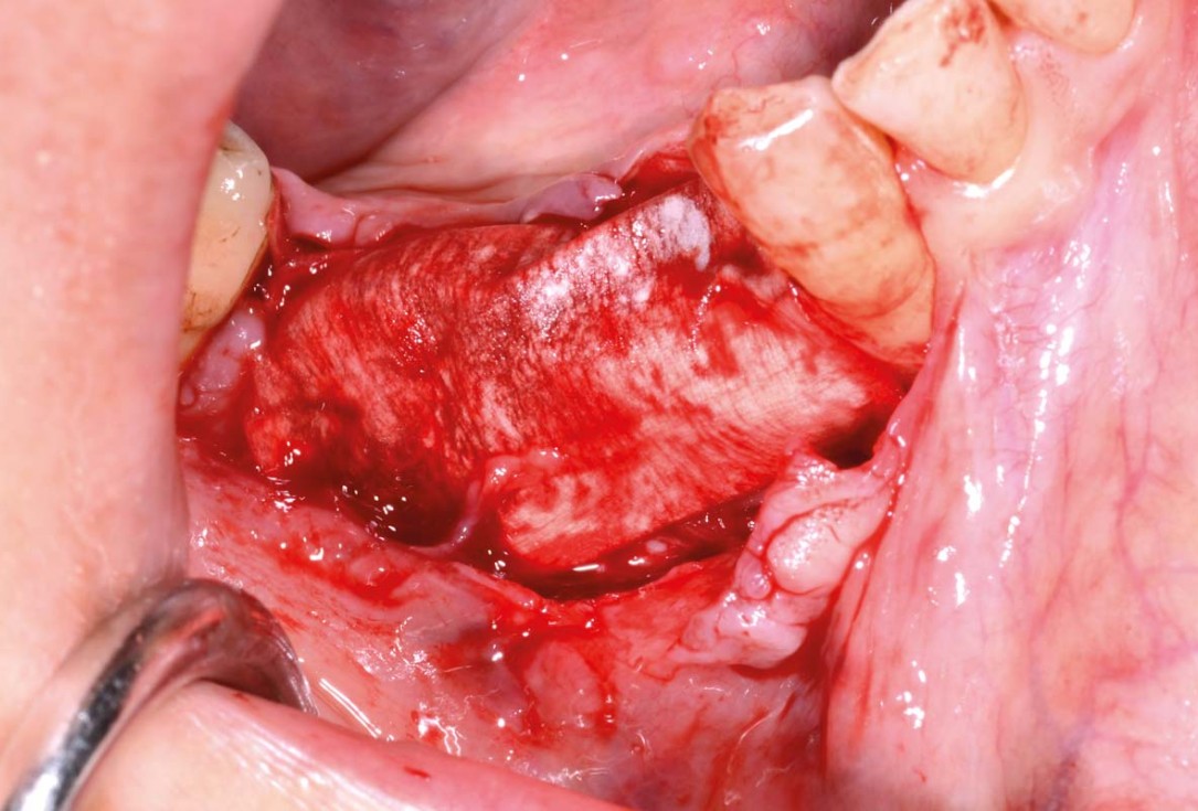
11/29 - Flap opening for removal of permamem®Vertical bone augmentation and broadening of attached gingiva using cerabone®, permamem® and mucoderm® - Dr. R. Naimoli
-
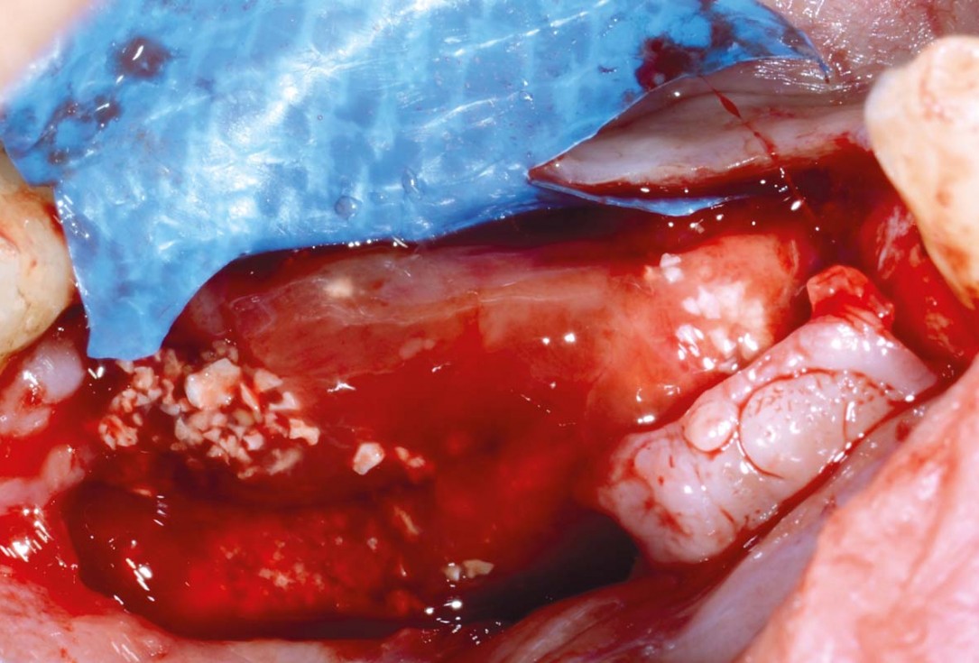
12/29 - Augmented area in maturation phase exposed under permamem®Vertical bone augmentation and broadening of attached gingiva using cerabone®, permamem® and mucoderm® - Dr. R. Naimoli
-
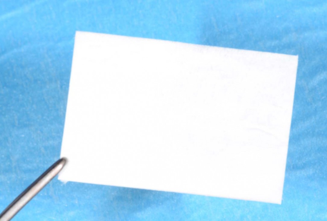
13/29 - Jason® membrane before hydrationVertical bone augmentation and broadening of attached gingiva using cerabone®, permamem® and mucoderm® - Dr. R. Naimoli
-
14/29 - Jason® membrane covering the augmented areaVertical bone augmentation and broadening of attached gingiva using cerabone®, permamem® and mucoderm® - Dr. R. Naimoli
-
15/29 - Flap closureVertical bone augmentation and broadening of attached gingiva using cerabone®, permamem® and mucoderm® - Dr. R. Naimoli
-
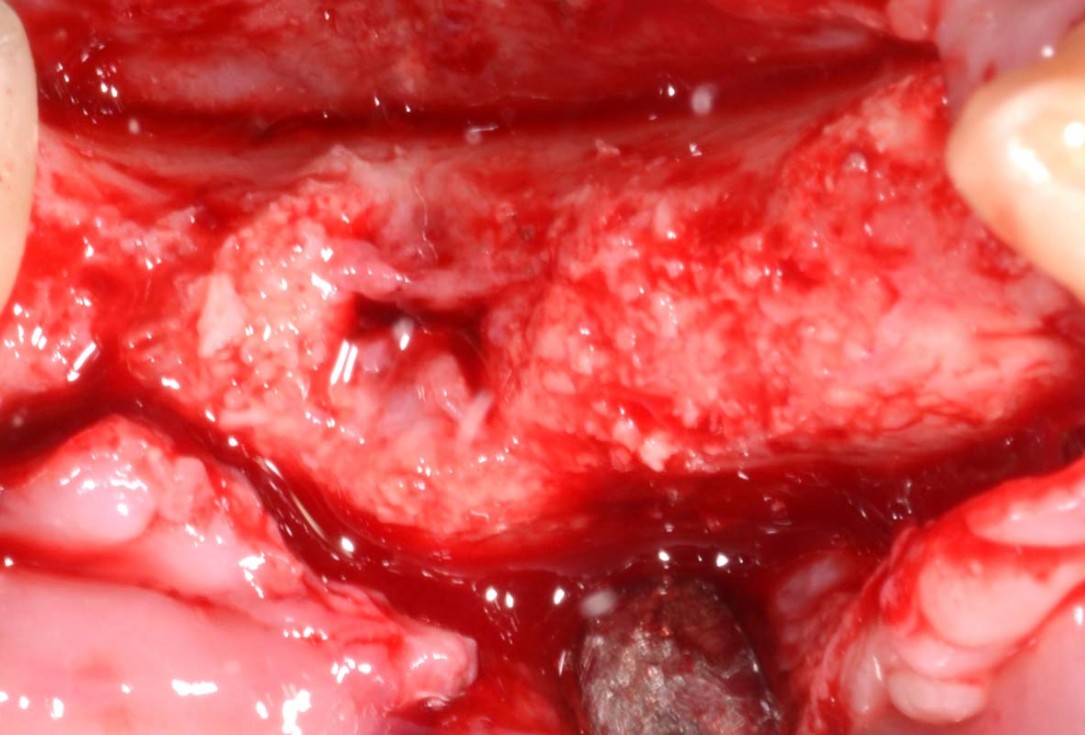
16/29 - Clinical view at re-entry, 8 months post-opVertical bone augmentation and broadening of attached gingiva using cerabone®, permamem® and mucoderm® - Dr. R. Naimoli
-
17/29 - Slight loss of the graft in regio 4.6, but still favorable for the insertion of the implantsVertical bone augmentation and broadening of attached gingiva using cerabone®, permamem® and mucoderm® - Dr. R. Naimoli
-
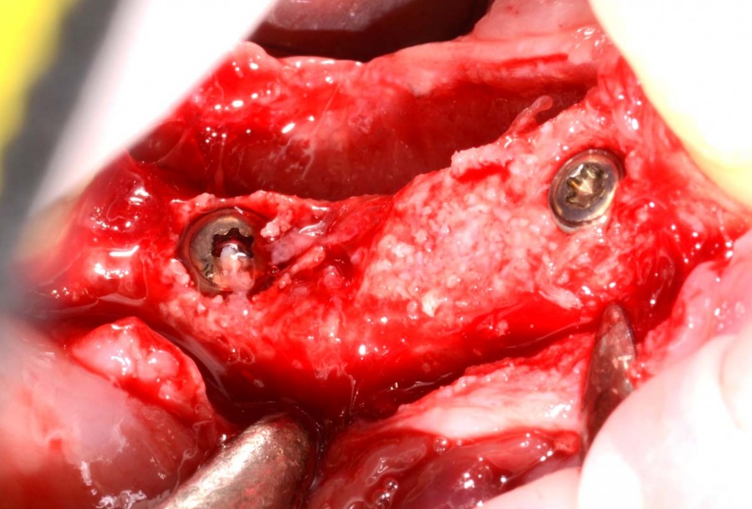
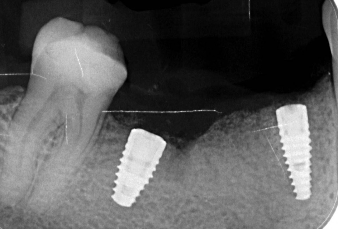
18/29 - Insertion of 2 Straumann BLT implants (regio 4.4: 3.3 x 10 mm, regio 4.6: 4.1x 8 mm), excellent primary stabilityVertical bone augmentation and broadening of attached gingiva using cerabone®, permamem® and mucoderm® - Dr. R. Naimoli
-
19/29 - Flap closureVertical bone augmentation and broadening of attached gingiva using cerabone®, permamem® and mucoderm® - Dr. R. Naimoli
-
20/29 - X-ray controlVertical bone augmentation and broadening of attached gingiva using cerabone®, permamem® and mucoderm® - Dr. R. Naimoli
-
21/29 - Clinical situation 3 months after implantationVertical bone augmentation and broadening of attached gingiva using cerabone®, permamem® and mucoderm® - Dr. R. Naimoli
-
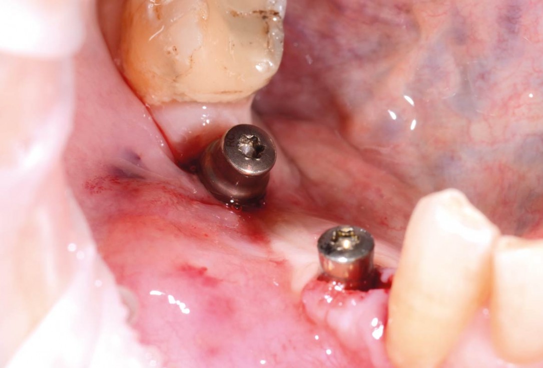
22/29 - Clinical view after insertion of the healing abutmentVertical bone augmentation and broadening of attached gingiva using cerabone®, permamem® and mucoderm® - Dr. R. Naimoli
-
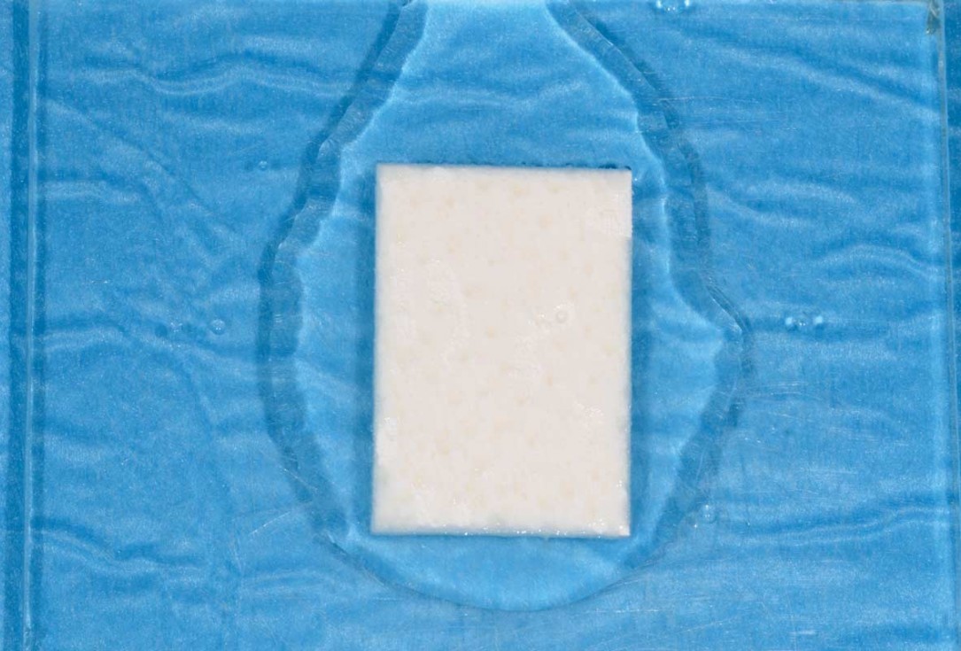
23/29 - Hydration of mucoderm®Vertical bone augmentation and broadening of attached gingiva using cerabone®, permamem® and mucoderm® - Dr. R. Naimoli
-
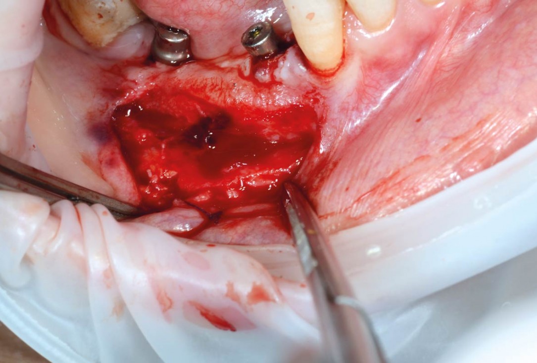
24/29 - Preparation of vestibular partial thickness flap (highlighting and isolation of mental nerve)Vertical bone augmentation and broadening of attached gingiva using cerabone®, permamem® and mucoderm® - Dr. R. Naimoli
-
25/29 - Fixation of mucoderm® to periosteum and flapVertical bone augmentation and broadening of attached gingiva using cerabone®, permamem® and mucoderm® - Dr. R. Naimoli
-
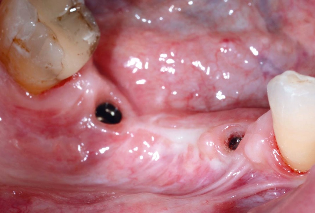
26/29 - Clinical view with broadened attached gingiva after 3 weeks, impression takingVertical bone augmentation and broadening of attached gingiva using cerabone®, permamem® and mucoderm® - Dr. R. Naimoli
-
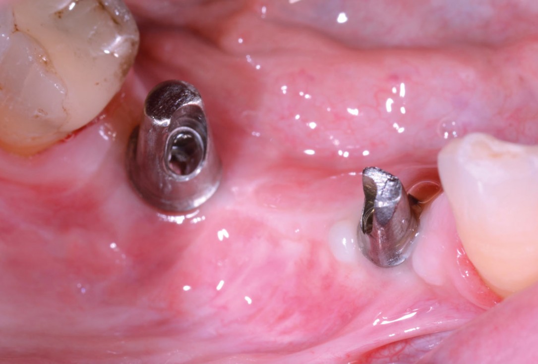
27/29 - Clinical view before installation of the fixed prosthetics, good amount of keratinized tissue around the implantsVertical bone augmentation and broadening of attached gingiva using cerabone®, permamem® and mucoderm® - Dr. R. Naimoli
-
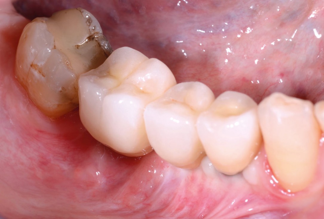
28/29 - Final prosthetic restorationVertical bone augmentation and broadening of attached gingiva using cerabone®, permamem® and mucoderm® - Dr. R. Naimoli
-
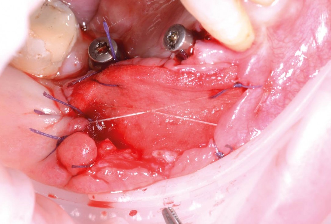
29/29 - Clinical view at control 3 month after final restorationVertical bone augmentation and broadening of attached gingiva using cerabone®, permamem® and mucoderm® - Dr. R. Naimoli

Initial clinical situation.

Initial clinical situation.

Initial clinical situation.

Baseline clinical situation.

Grafting of the extraction socket with small cerabone® granules.

Situation before tooth extraction

Situation after tooth extraction.

Initial situation - A young female 34 years old lost her front teeth in an surfing accident and she had a 5 unit bridge supported by her upper left lateral and right canine. The restoration failed and both supporting crowns have exposed and leaking margins.

Tooth 16 furcation involvement with gingival marginal recession and large Class 5 filling

Pre-surgical situation.

Initial clinical situation.

Initial clinical situation showing bone wall defect.

Pre-operative situation. Lateral view.

Situation before tooth extraction.

Situation after tooth extraction.

Initial clinical situation with pronounced vertical and horizontal bone defect

Initial panoramic x-ray with failing tooth 16

Implant placed in the deficient site. permamem® in place for covering.

Initial clinical situation. Atrophic maxillary ridge.

Intra-operative view.

Extraction socket with bone wall defect

Immediately placed implant covered with permamem®. permamem® passively immobilized by sutures and intentionally left exposed to the oral cavity.

Baseline clinical situation.
