Treatment of a combined horizontal and vertical bone defect in the maxilla with maxgraft® cortico in the allogenic shell technique - Dr. R. Würdinger
-
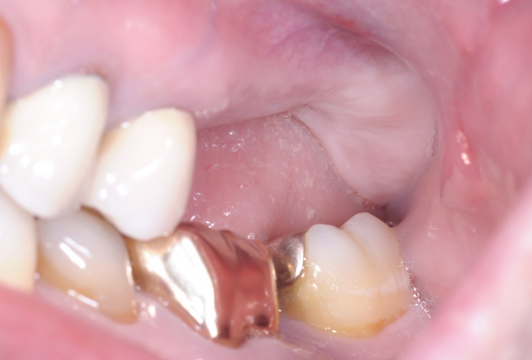
1/15 - Clinical situationTreatment of a combined horizontal and vertical bone defect in the maxilla with maxgraft® cortico in the allogenic shell technique - Dr. R. Würdinger
-
2/15 - Preoperative OPG displaying the vertical bone defectTreatment of a combined horizontal and vertical bone defect in the maxilla with maxgraft® cortico in the allogenic shell technique - Dr. R. Würdinger
-
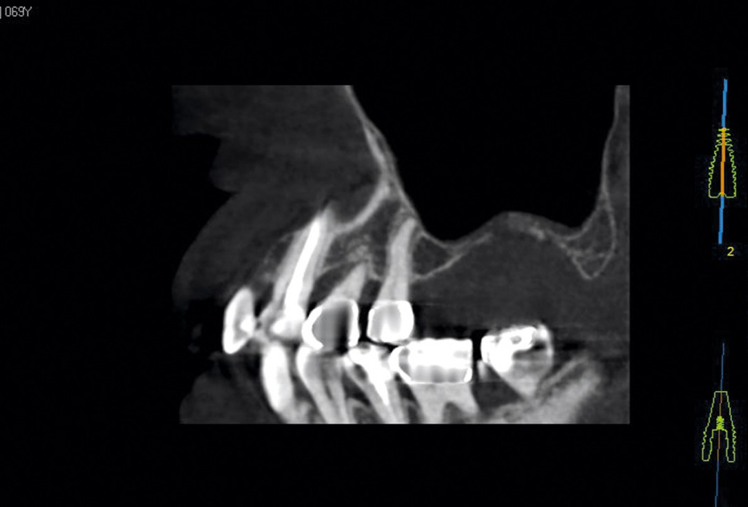
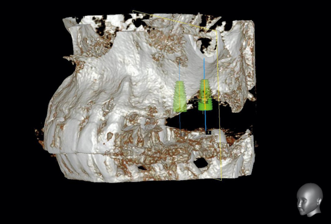
3/15 - Preoperative OPG - planning of the implant placementTreatment of a combined horizontal and vertical bone defect in the maxilla with maxgraft® cortico in the allogenic shell technique - Dr. R. Würdinger
-
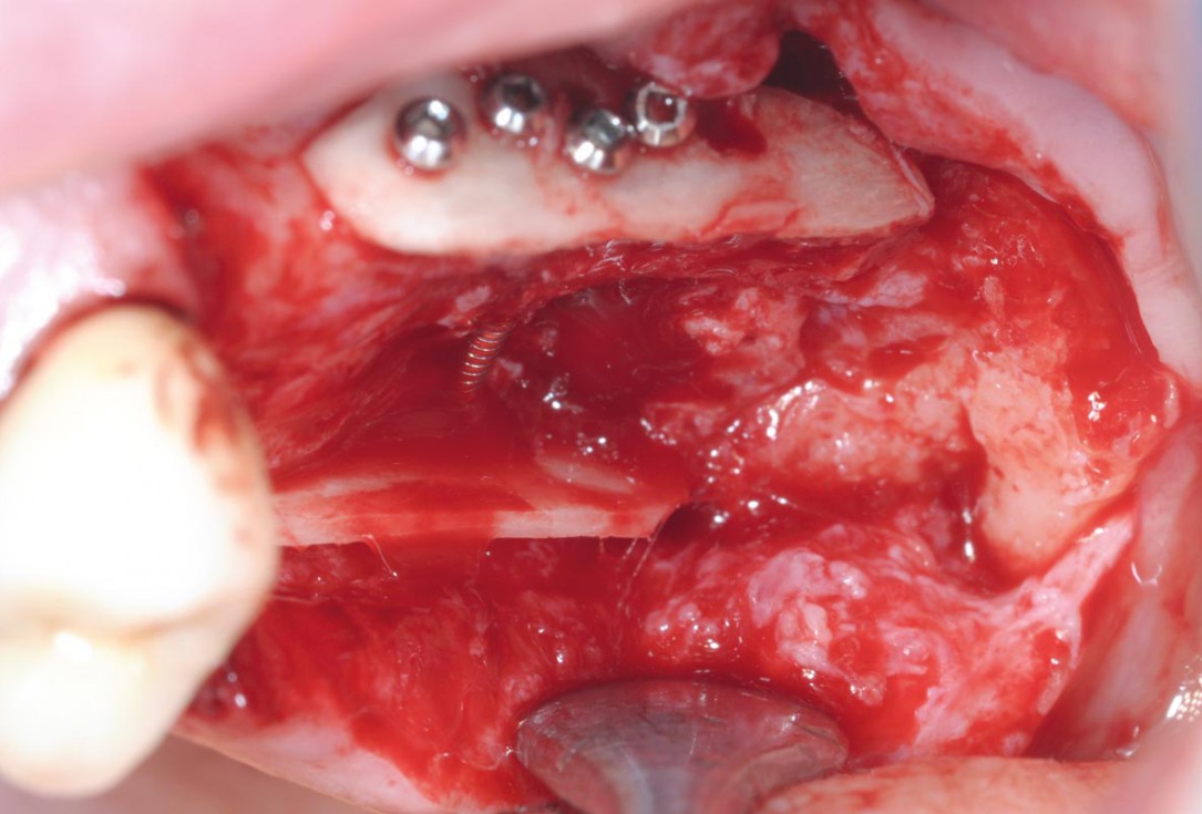
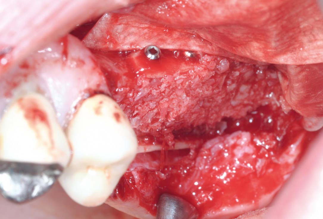
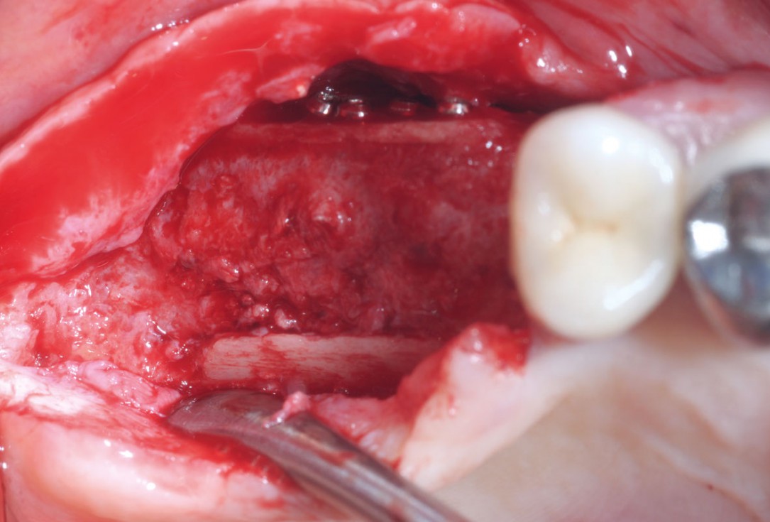
4/15 - Intraoperative view after fixation of maxgraft® cortico with a micro screw system. Placement in parallel technique to obtain a horizontal and vertical augmentationTreatment of a combined horizontal and vertical bone defect in the maxilla with maxgraft® cortico in the allogenic shell technique - Dr. R. Würdinger
-
5/15 - Filling of the container with maxgraft® cortico-cancellous granulesTreatment of a combined horizontal and vertical bone defect in the maxilla with maxgraft® cortico in the allogenic shell technique - Dr. R. Würdinger
-
6/15 - Augmented site covered with Jason® membraneTreatment of a combined horizontal and vertical bone defect in the maxilla with maxgraft® cortico in the allogenic shell technique - Dr. R. Würdinger
-
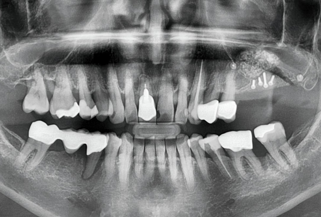
7/15 - X-ray control after augmentationTreatment of a combined horizontal and vertical bone defect in the maxilla with maxgraft® cortico in the allogenic shell technique - Dr. R. Würdinger
-
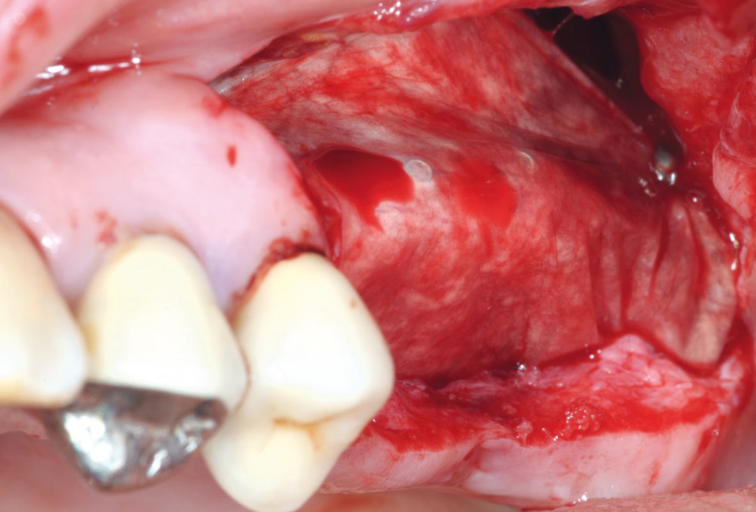
8/15 - Clinical situation five months after augmentation, time of re-entryTreatment of a combined horizontal and vertical bone defect in the maxilla with maxgraft® cortico in the allogenic shell technique - Dr. R. Würdinger
-
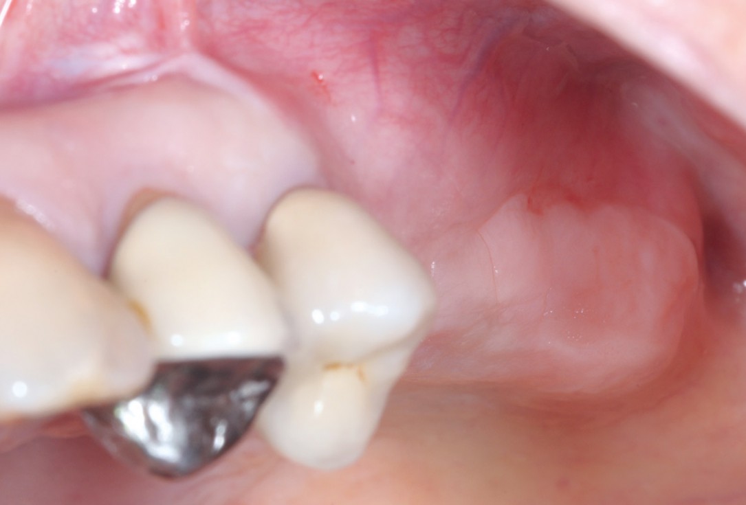
9/15 - Occlusal view of the remodeled augmented site at re-entryTreatment of a combined horizontal and vertical bone defect in the maxilla with maxgraft® cortico in the allogenic shell technique - Dr. R. Würdinger
-
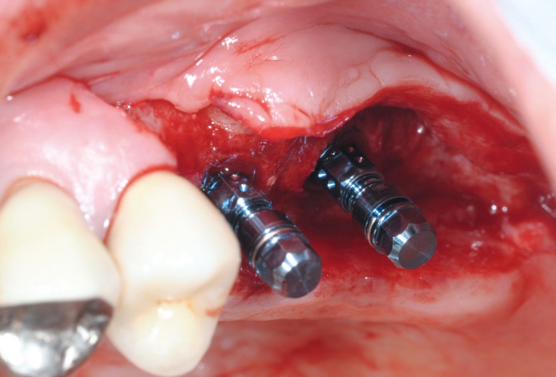
10/15 - Insertion of two Straumann Bone Level Tapered implants 4.8, 10 mm, lateral viewTreatment of a combined horizontal and vertical bone defect in the maxilla with maxgraft® cortico in the allogenic shell technique - Dr. R. Würdinger
-
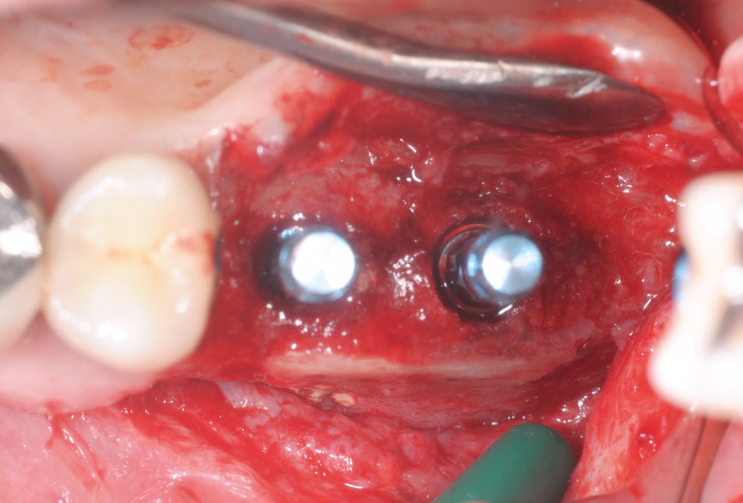
11/15 - Occlusal viewTreatment of a combined horizontal and vertical bone defect in the maxilla with maxgraft® cortico in the allogenic shell technique - Dr. R. Würdinger
-
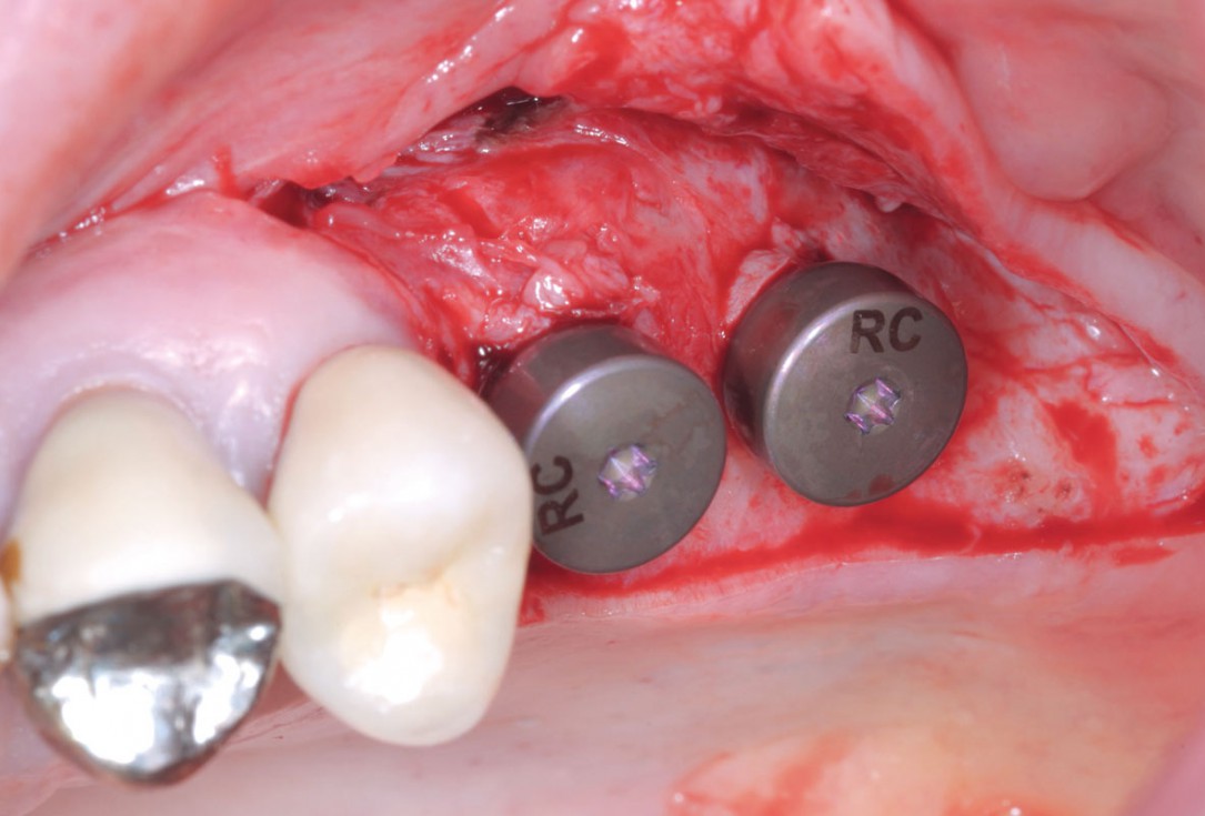
12/15 - Placement of gingiva abutmentTreatment of a combined horizontal and vertical bone defect in the maxilla with maxgraft® cortico in the allogenic shell technique - Dr. R. Würdinger
-
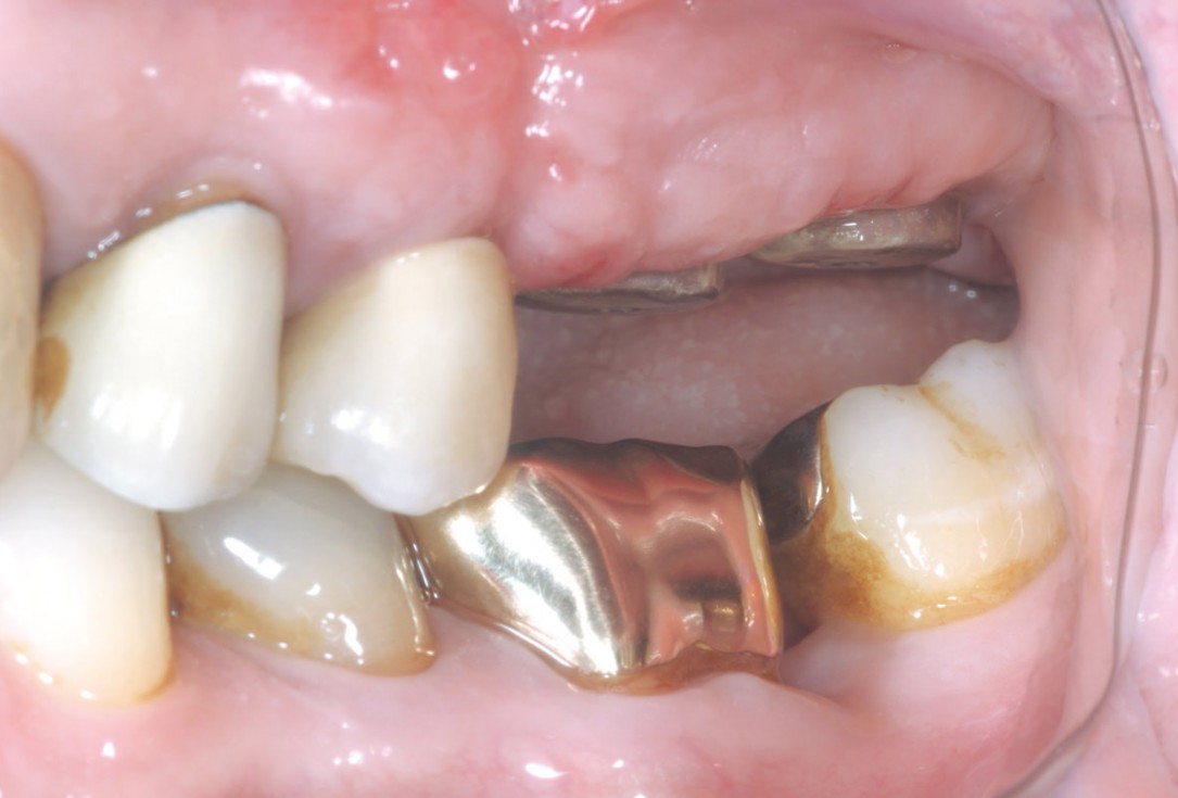
13/15 - Clinical situation 14 days after implant uncoveringTreatment of a combined horizontal and vertical bone defect in the maxilla with maxgraft® cortico in the allogenic shell technique - Dr. R. Würdinger
-
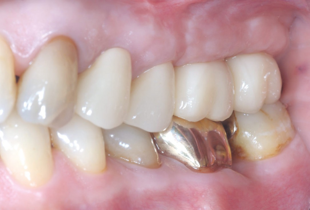
14/15 - Final prosthetic restoration- proper implant-crown ratio compared to the neighboring teethTreatment of a combined horizontal and vertical bone defect in the maxilla with maxgraft® cortico in the allogenic shell technique - Dr. R. Würdinger
-
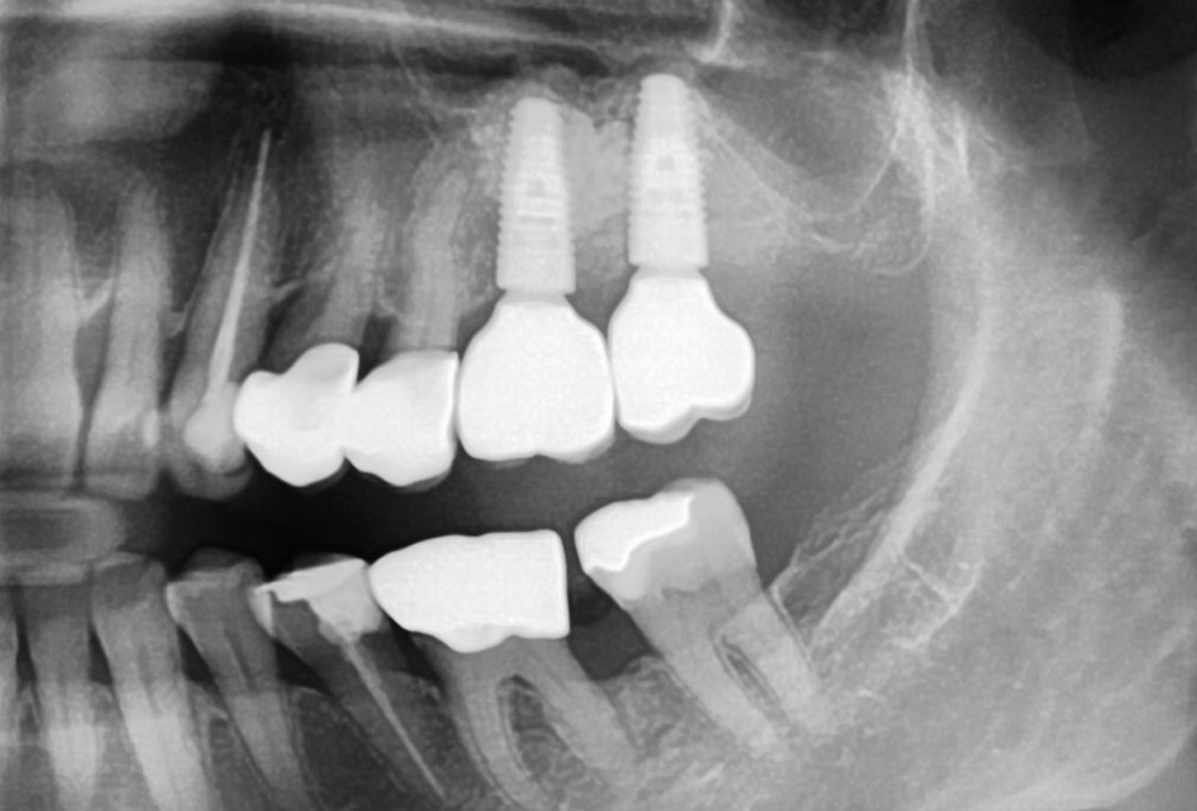
15/15 - X-ray control after final prosthetic restoration six months after augmentationTreatment of a combined horizontal and vertical bone defect in the maxilla with maxgraft® cortico in the allogenic shell technique - Dr. R. Würdinger

Baseline clinical situation.

Situation before extraction of the teeth

Initial clinical situation

Initial situation after extraction of tooth 21 after 6 months

Preparation of a single tooth defect with severely resorbed vestibular wall

Initial clinical situation.

Initial clinical situation

Tooth 16 furcation involvement with gingival marginal recession and large Class 5 filling

Initial x-ray, ten years post implantationem alio loco, large peri-implant bone loss

Extraction socket with bone wall defect

Situation before tooth extraction

Initial clinical situation

Initial X-ray presenting a very deep intrabony defect of tooth 21

Implant placed in the deficient site. permamem® in place for covering.

Initial situation – Treatment plan: Replace the adhesive upper left central incisor bridge with a dental implant

Occlusal view of attached maxgraft® cortico at the buccal site

Initial x-ray, tooth 25 compromised and to be extracted

Clinical situation at baseline: Situation after tooth extraction UR1 due to a failed endodontic treatment 3 months previously

Alveolar socket before soft and hard tissue augmentation

Pre-operative situation; tooth 21 proved not to be worth preserving

Initial clinical situation: 9 mm pocket depth associated with root fracture

Initial situation - A young female 34 years old lost her front teeth in an surfing accident and she had a 5 unit bridge supported by her upper left lateral and right canine. The restoration failed and both supporting crowns have exposed and leaking margins.

Initial situation - endodontically failing tooth 22, very thin biotype, high lip line and esthetic expectations

Preoperative x-ray, severe bone atrophy

Initial situation - broken and missing upper right central incisor (UR1). This tooth was removed long time ago and there were signs of bone loss and resorption due to the bone remodelling. Patient was also undergoing orthodontic treatment due to the loss of mesio-distal space.
