Multiple gingival recessions treated with the modified coronally advanced tunnel in conjunction with Straumann® Emdogain® and autologous CTG - Dr. R. Cosgarea
-
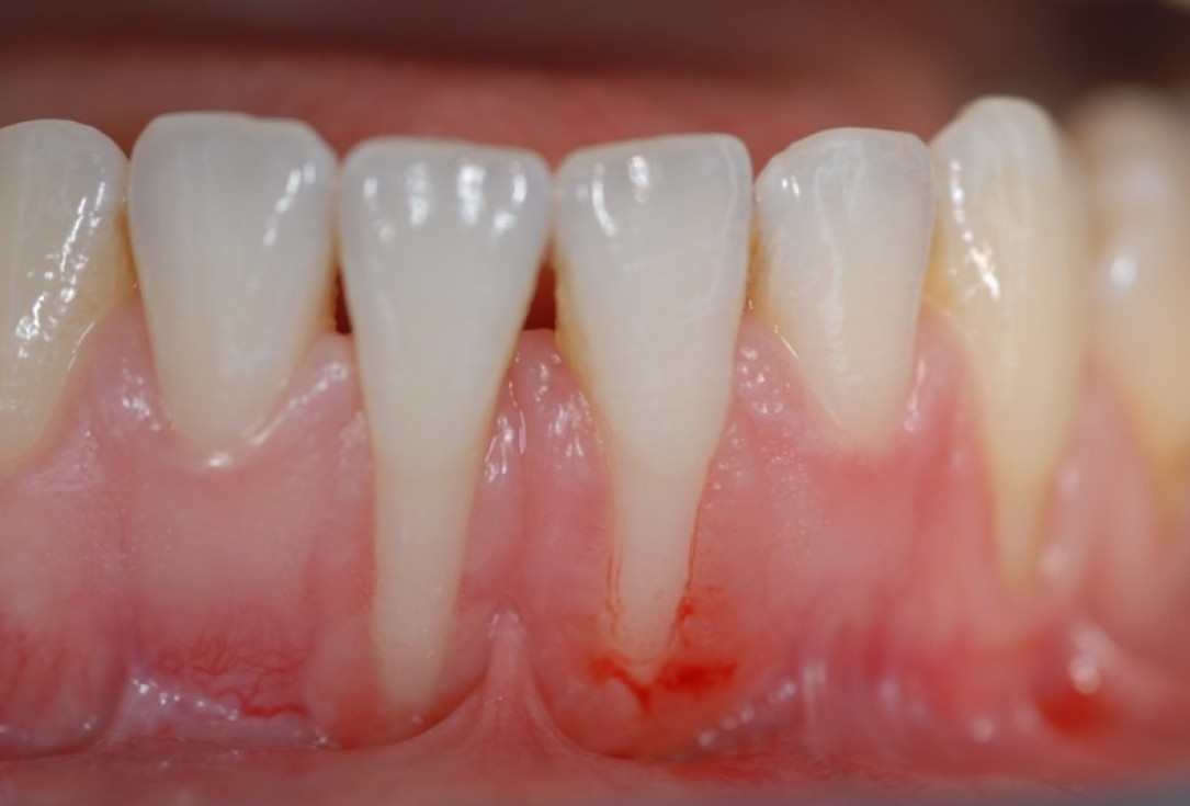
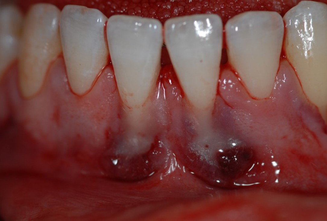
01/10 - Pre-operative clinical view.Multiple gingival recessions treated with the modified coronally advanced tunnel in conjunction with Straumann® Emdogain® and autologous CTG - Dr. R. Cosgarea
-
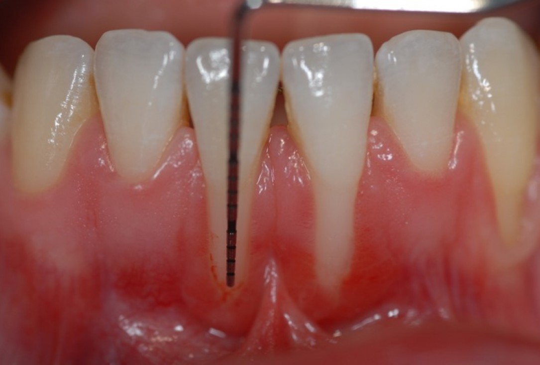
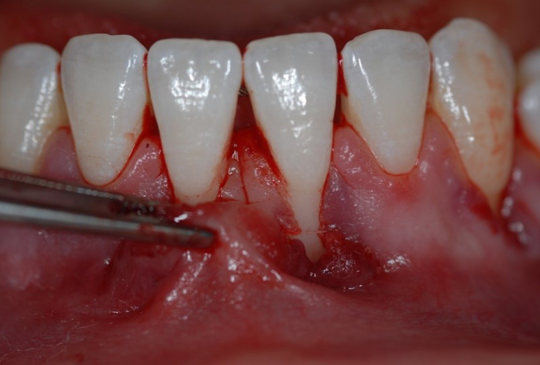
02/10 - Recession depth of 9 mm at teeth 31 and 41.Multiple gingival recessions treated with the modified coronally advanced tunnel in conjunction with Straumann® Emdogain® and autologous CTG - Dr. R. Cosgarea
-
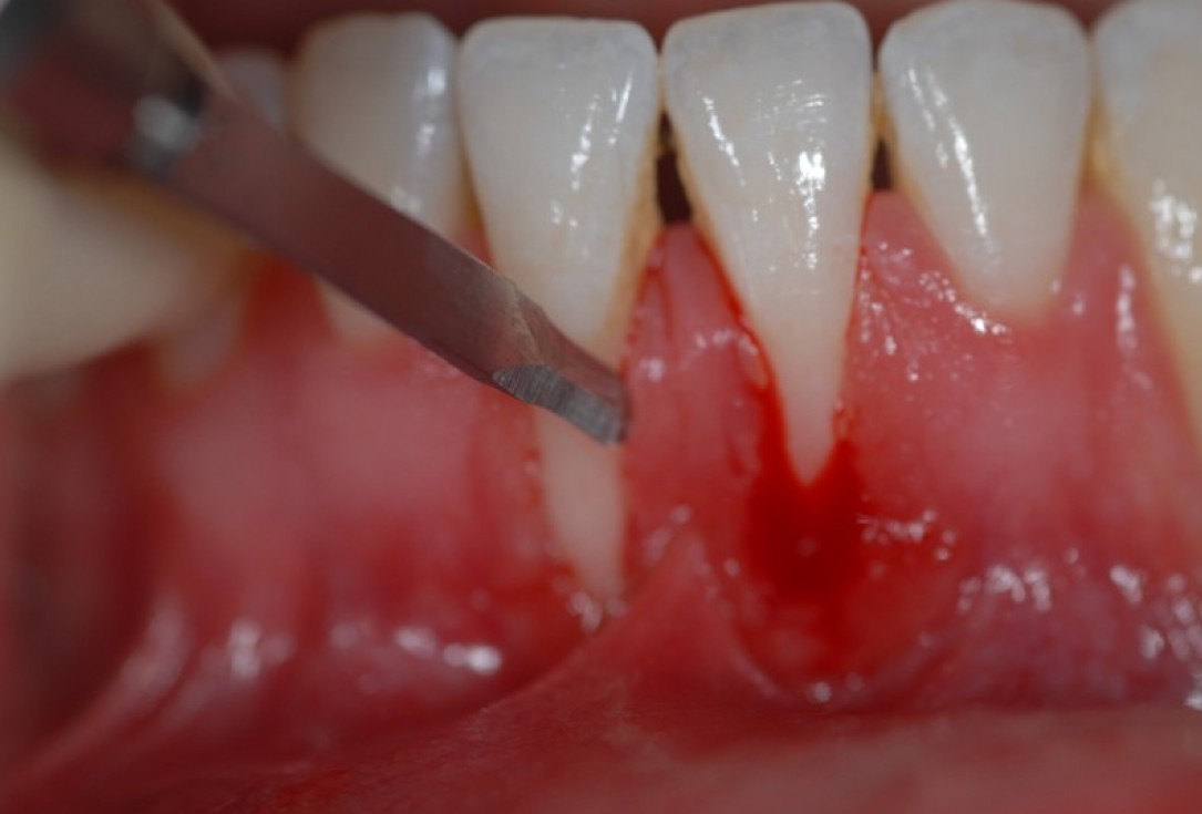
03/10 - Intrasulcular incision.Multiple gingival recessions treated with the modified coronally advanced tunnel in conjunction with Straumann® Emdogain® and autologous CTG - Dr. R. Cosgarea
-
04/10 - Preparation of the tunnel using tunneling instruments.Multiple gingival recessions treated with the modified coronally advanced tunnel in conjunction with Straumann® Emdogain® and autologous CTG - Dr. R. Cosgarea
-
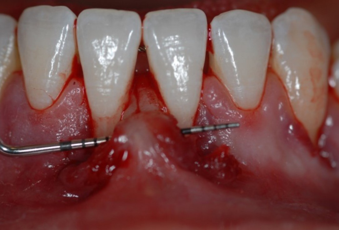
05/10 - Test for tension-free preparation of the tunnel.Multiple gingival recessions treated with the modified coronally advanced tunnel in conjunction with Straumann® Emdogain® and autologous CTG - Dr. R. Cosgarea
-
06/10 - Application of Straumann® PrefGel® for 2 minutes onto the root surfaces.Multiple gingival recessions treated with the modified coronally advanced tunnel in conjunction with Straumann® Emdogain® and autologous CTG - Dr. R. Cosgarea
-
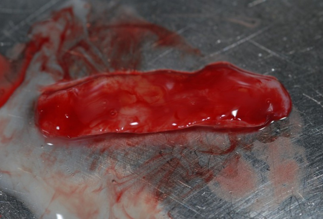
07/10 - Coating of the autologous CTG with Straumann® Emdogain® for 5 minutes.Multiple gingival recessions treated with the modified coronally advanced tunnel in conjunction with Straumann® Emdogain® and autologous CTG - Dr. R. Cosgarea
-
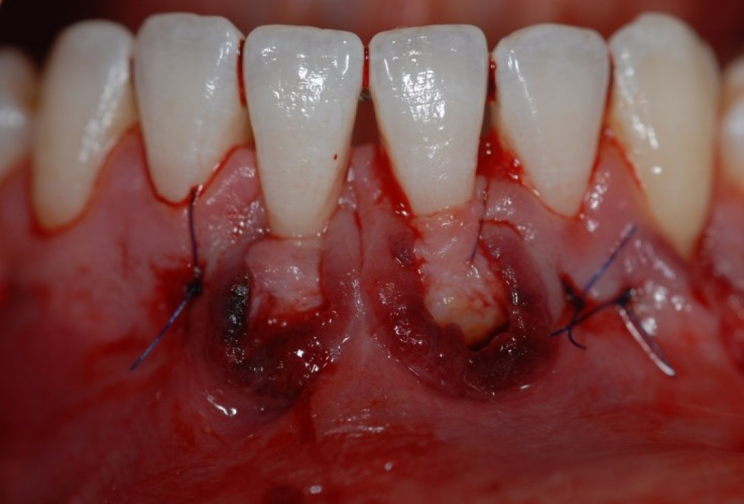
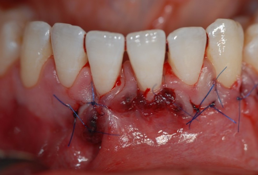
08/10 - After rinsing and application of Straumann® Emdogain® onto the root surfaces, insertion of the CTG into the tunnel and fixation with sutures.Multiple gingival recessions treated with the modified coronally advanced tunnel in conjunction with Straumann® Emdogain® and autologous CTG - Dr. R. Cosgarea
-
09/10 - Coverage of the CTG and the recession by coronal and lateral movement of the tunnel. Fixation by sling sutures.Multiple gingival recessions treated with the modified coronally advanced tunnel in conjunction with Straumann® Emdogain® and autologous CTG - Dr. R. Cosgarea
-
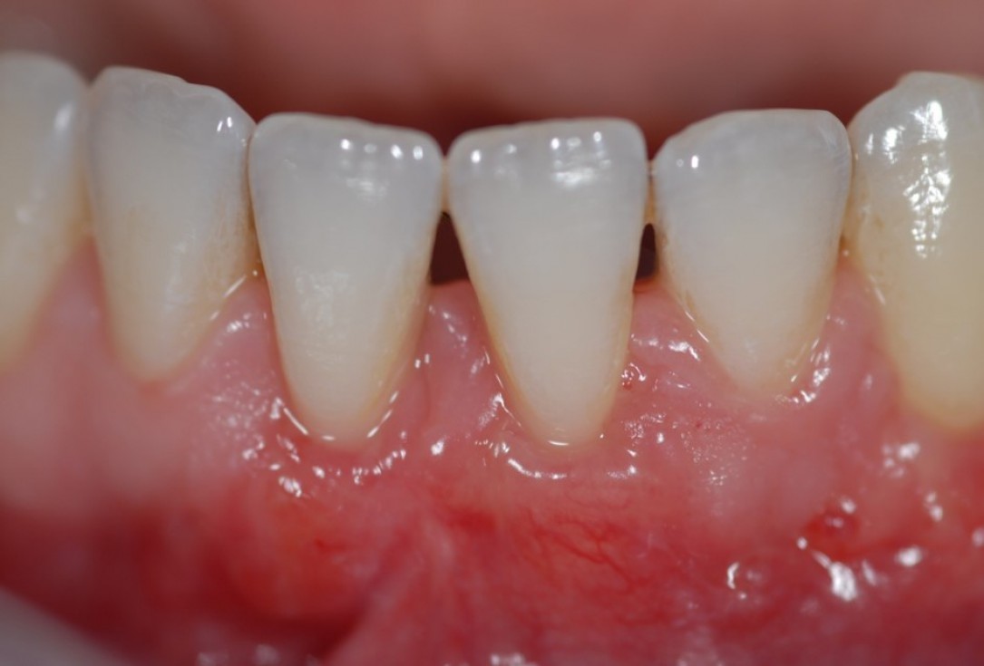
10/10 - Clinical situation 3 weeks post-operative.Multiple gingival recessions treated with the modified coronally advanced tunnel in conjunction with Straumann® Emdogain® and autologous CTG - Dr. R. Cosgarea

Alveolar socket before soft and hard tissue augmentation

Pre-operative OPG shows deep vertical intrabony defects on the distal aspects of teeth 13 and 14.

Radiographic view before periodontal regenerative therapy with Straumann® Emdogain®. A deep intrabony defect appeared mesially and distally on the left mandibular first premolar. Pre-surgical probing measured 8 mm. The defect morphology presented as well-contained.

Baseline clincial situation and pre-surgical probing.

Pre-operative radiographic view.

Baseline clinical situation. Recession depth of 6 mm at tooth 31.

Initial clinical situation

Pre-operative X-ray. Hopless tooth 21.

Pre-operative clinical situation.

Pre-operative radiograph. Intrabony defect on the mesial aspect of tooth 14.

Pre-operative clinical situation. Gingival recessions at teeth 11 and 21.

Pre-surgical situation. Multiple adjacent gingival recessions at teeth 12, 13 and 14.

Pre-operative clinical view. Multiple adjacent gingival recessions.

Initial situation: bone loss due to lack of physical load of bridge retained region 11

Pre-operative radiographic view. Intrabony defect on the distal aspect of the lateral incisor.

Situation after tooth removal.

Pre-operative clinical situation.

Pre-surgical clinical situation, buccal view.

Pre-operative probing pocket depth (PPD) at the distal aspect of tooth 11 was 7 mm.

Pre-surgical clinical situation. Deep gingival recessions at both upper canine.

Pre-operative clinical situation. Multiple adjacent gingival recessions.

Pre-operative clinical situation. Shallow multiple adjacent gingival recessions in the first quadrant.

Initial situation: 40 year old female patient with extensive scar tissue after several surgeries restored with a Rochette bridge
