Horizontal ridge augmentation with maxgraft® cortico - M.Sc. E. Kapogianni
-
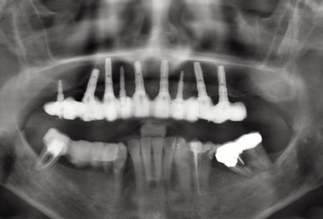
01 / 20 - OPG of the initial situation – provision of missing denture in regio 44 to 47 by a resin-retained bridgeHorizontal ridge augmentation with maxgraft® cortico - M.Sc. E. Kapogianni
-
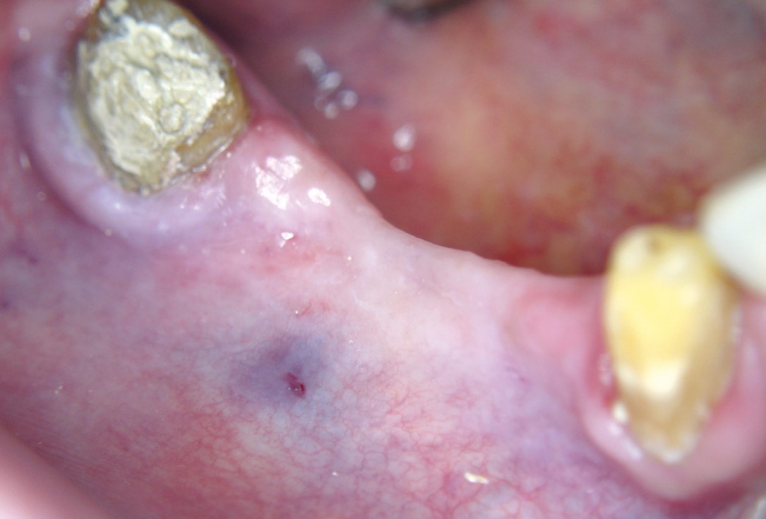
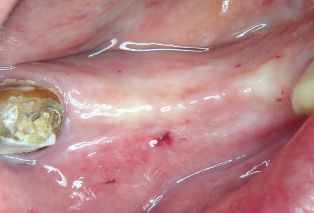
02 / 20 - Thin soft tissue and reduced mandibular widthHorizontal ridge augmentation with maxgraft® cortico - M.Sc. E. Kapogianni
-
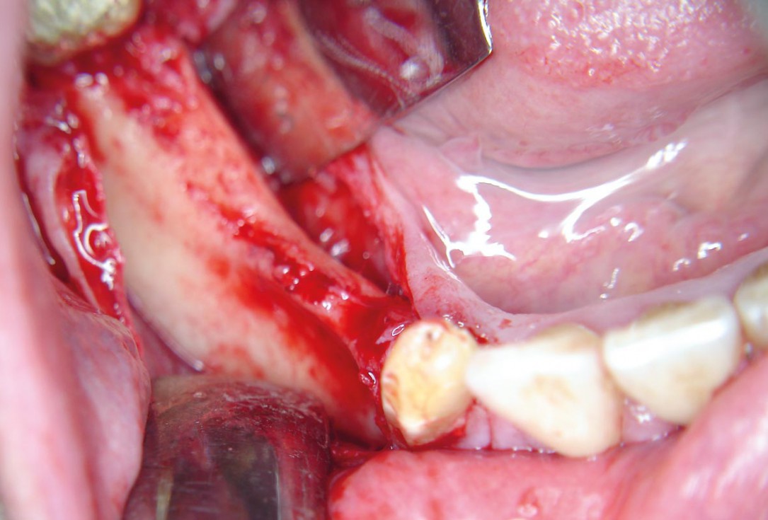
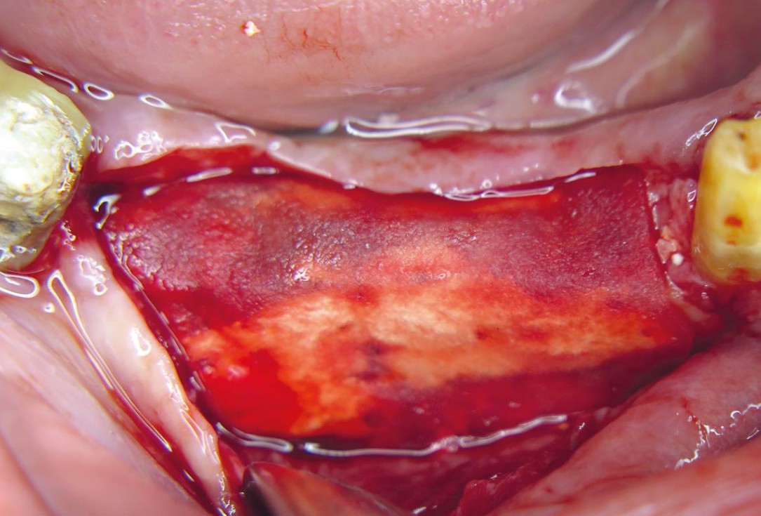
03 / 20 - Flap projection shows pronounced bone lossHorizontal ridge augmentation with maxgraft® cortico - M.Sc. E. Kapogianni
-
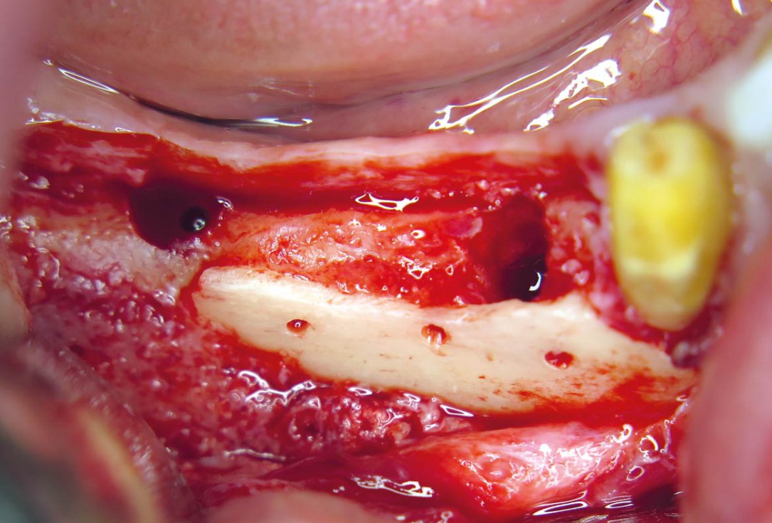
04 / 20 - Decortication of the host bone and fixation of a defect-adapted cortical plateHorizontal ridge augmentation with maxgraft® cortico - M.Sc. E. Kapogianni
-
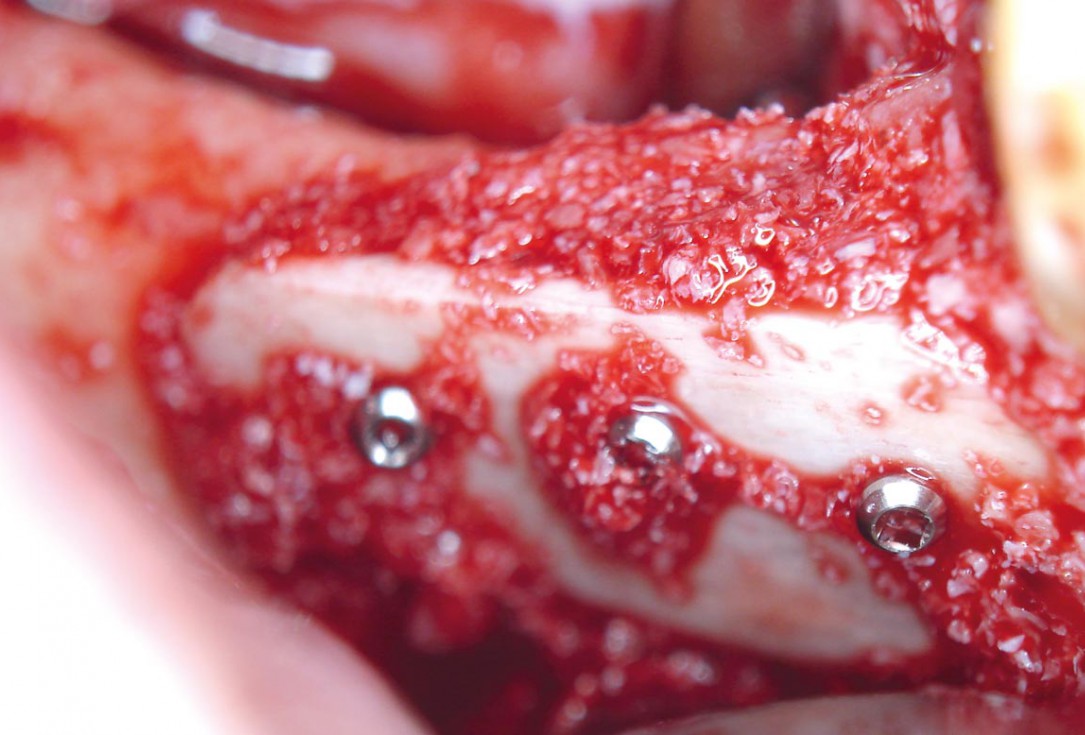
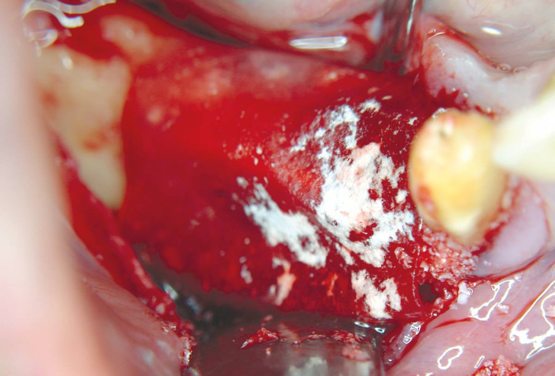
05 / 20 - Defect-filling and contouring with cancellous allogenic bone chipsHorizontal ridge augmentation with maxgraft® cortico - M.Sc. E. Kapogianni
-
06 / 20 - Augmentation site covering by application of a collprotect® membraneHorizontal ridge augmentation with maxgraft® cortico - M.Sc. E. Kapogianni
-
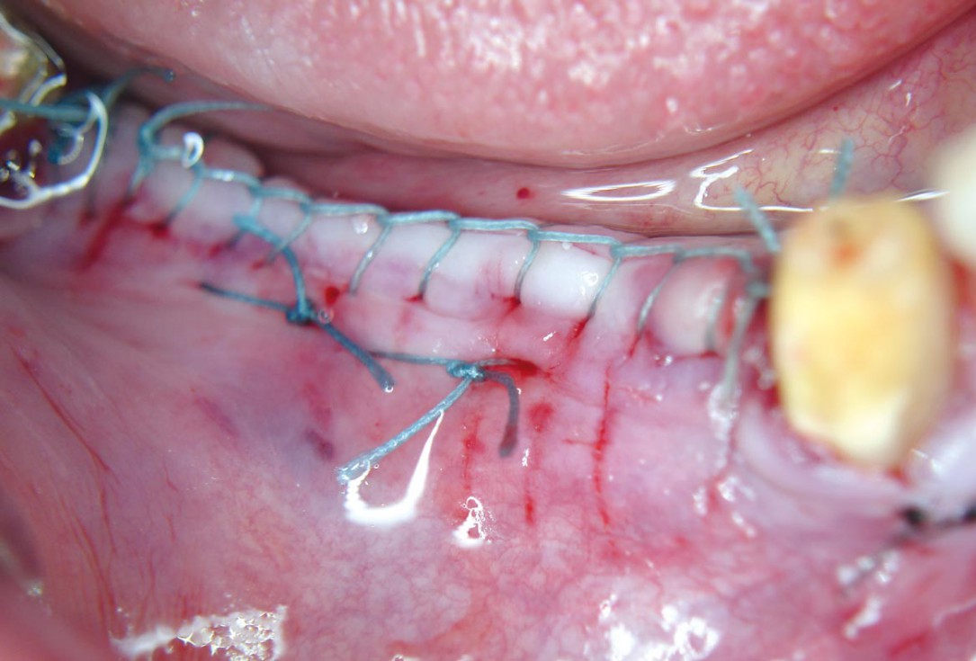
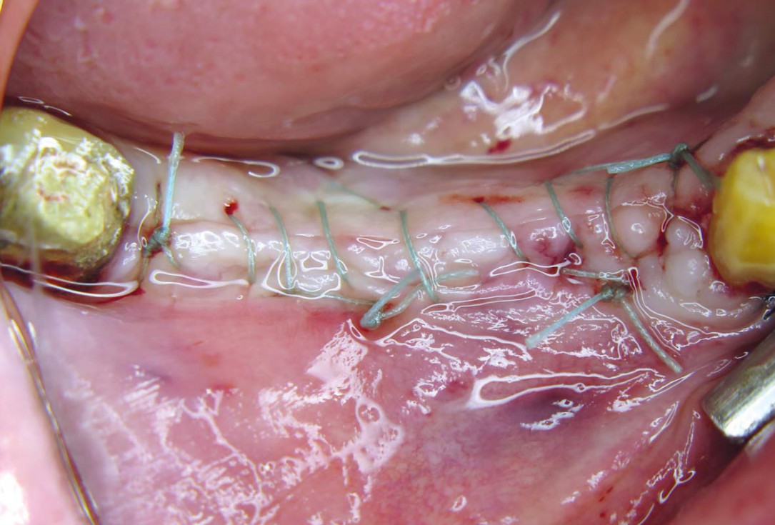
07 / 20 - Tension-free wound closure without compression on the augmentation siteHorizontal ridge augmentation with maxgraft® cortico - M.Sc. E. Kapogianni
-
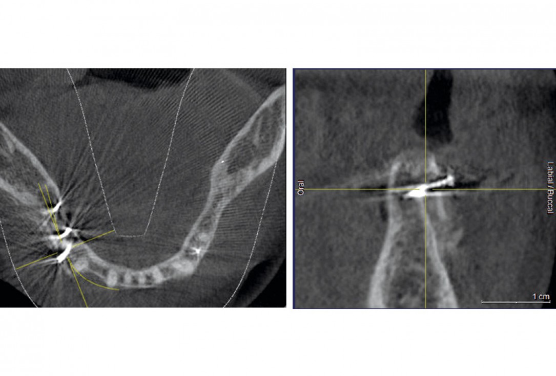
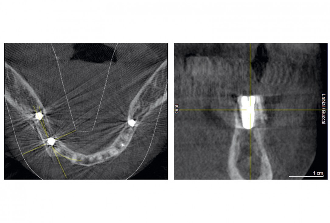
08 / 20 - CBCT recording after the augmentation shows the cortical plate about 3 to 4 mm distant from the host boneHorizontal ridge augmentation with maxgraft® cortico - M.Sc. E. Kapogianni
-
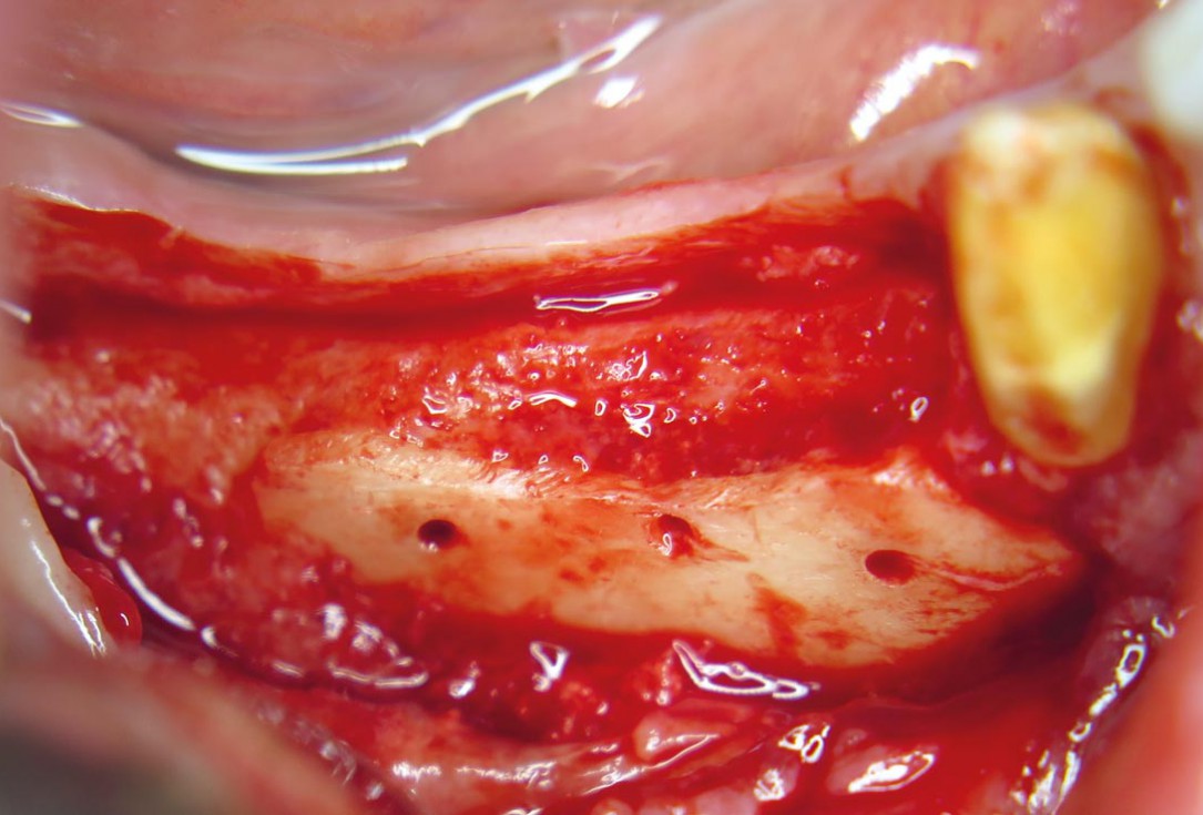
09 / 20 - Reentry after 5 months of healing shows excellent bone regenerationHorizontal ridge augmentation with maxgraft® cortico - M.Sc. E. Kapogianni
-
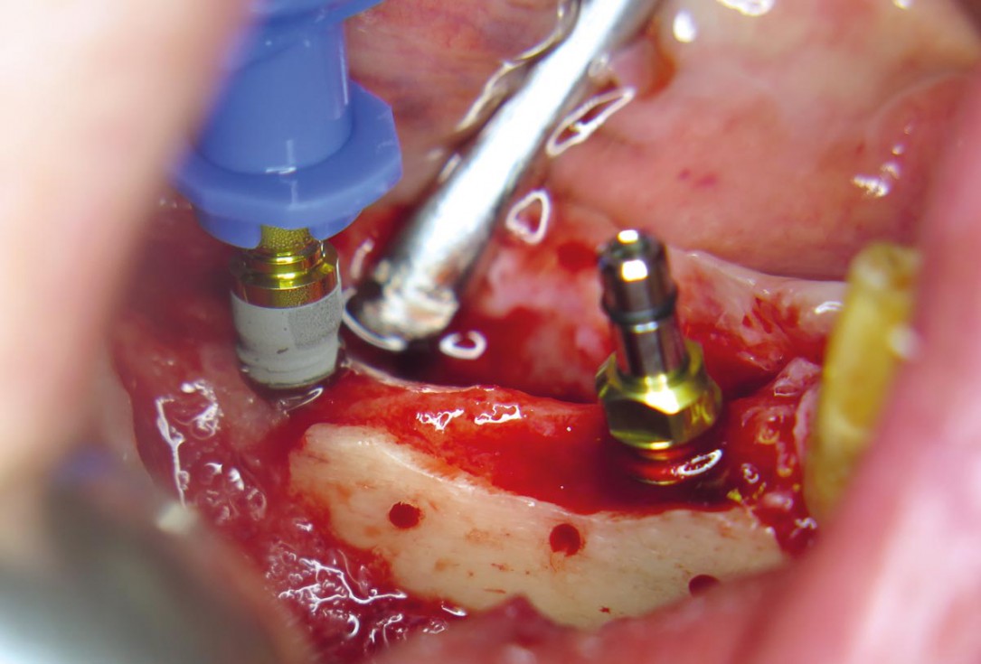
10 / 20 - Placing of pilot drills into the new formed bone tissueHorizontal ridge augmentation with maxgraft® cortico - M.Sc. E. Kapogianni
-
11 / 20 - Insertion of two dental implants in regio 44 and 46Horizontal ridge augmentation with maxgraft® cortico - M.Sc. E. Kapogianni
-
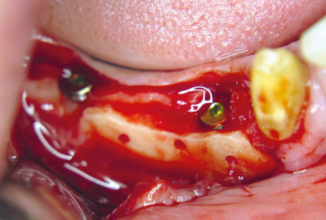
12 / 20 - Stable and fully submerged positioning of implantsHorizontal ridge augmentation with maxgraft® cortico - M.Sc. E. Kapogianni
-
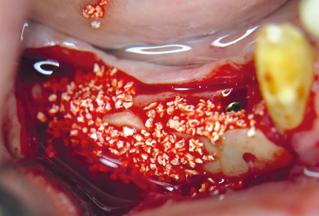
13 / 20 - Contouring with cerabone® for optimal volume stability and an aesthetic outcomeHorizontal ridge augmentation with maxgraft® cortico - M.Sc. E. Kapogianni
-
14 / 20 - Surgical site covering with a collprotect® membraneHorizontal ridge augmentation with maxgraft® cortico - M.Sc. E. Kapogianni
-
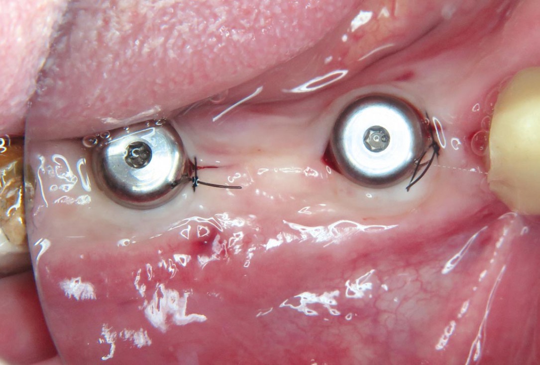
15 / 20 - Tension-and compression-free suturesHorizontal ridge augmentation with maxgraft® cortico - M.Sc. E. Kapogianni
-
16 / 20 - CBCT after the implantation shows position of the implant within vital bone tissue and the adjacent allogenic cortical plateHorizontal ridge augmentation with maxgraft® cortico - M.Sc. E. Kapogianni
-
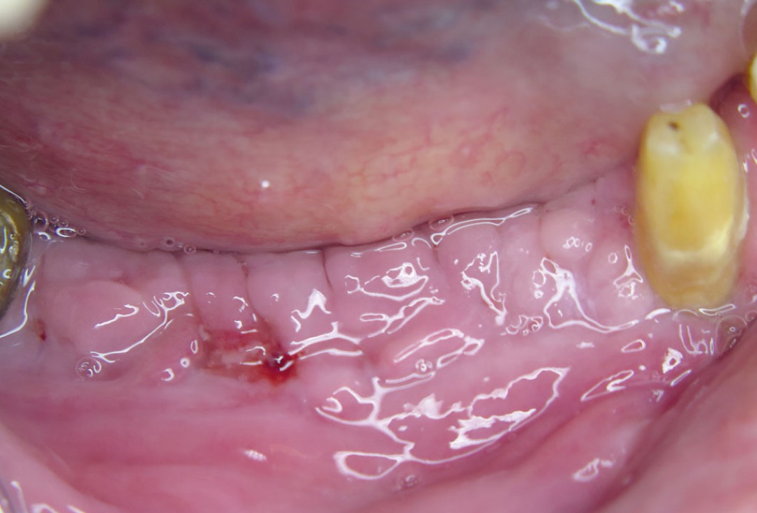
17 / 20 - Soft tissue healing one week after implantationHorizontal ridge augmentation with maxgraft® cortico - M.Sc. E. Kapogianni
-
18 / 20 - Excellent soft tissue situation after three months of healingHorizontal ridge augmentation with maxgraft® cortico - M.Sc. E. Kapogianni
-
19 / 20 - Placing of gingiva formers for an aesthetic margin between the final crown and the soft tissueHorizontal ridge augmentation with maxgraft® cortico - M.Sc. E. Kapogianni
-
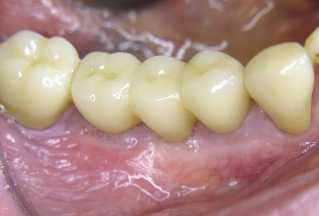
20 / 20 - Final implant-retained denture with natural appearance one month after placing of gingiva formersHorizontal ridge augmentation with maxgraft® cortico - M.Sc. E. Kapogianni

Initial clinical situation. Atrophic maxillary ridge.

Initial x-ray showing bone loss around implants placed 5 years ago in another dental clinic

Initial view of the case. Discoloration of 1.1 and mild class I gingival recession

Situation after tooth removal.

Initial clinical situation with gum recession and labial bone loss eight weeks following tooth extraction

Three implants placed in a narrow posterior mandible

Pre-operative clinical situation.

Clinical situation with narrow alveolar ridge in the lower jaw

Initial clinical situation showing bone wall defect.

Initial clinical situation.

Pre-surgical situation.

Initial situation: missing teeth #11 & 12 and badly broken #21 root

Pre-operative OPG shows deep vertical intrabony defects on the distal aspects of teeth 13 and 14.

Instable bridge situation with abscess formation at tooth #15 after apicoectomy

Initial clinical situation.

Implant insertion in atrophic alveolar ridge

Preoperative clinical situation

Pre-operative OPG

Pre-operative X-ray. Hopless tooth 21.

Pre-surgical situation. Teeth 26 and 27 missing.

Extraction of tooth 21 after endodontic treatment

Pre-surgical probing reveals a deep intrabony defect on the distal aspect of the upper canine.

Initial clinical situation with single tooth gap in regio 21

Pre-operative radiographic view. Intrabony defect on the distal aspect of the lateral incisor.

Clinical situation before extraction and implantation

Pre-operative radiographic view.
