Circular bone splitting with maxresorb® & collprotect® membrane - PD Dr. J. Neugebauer
-
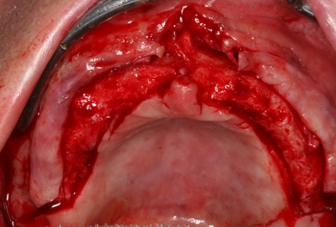
01/10 - Surgical presentation of the alveolar ridge with reduced amount of horizontal bone availableCircular bone splitting with maxresorb® & collprotect® membrane - PD Dr. J. Neugebauer
-
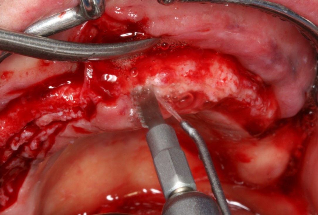
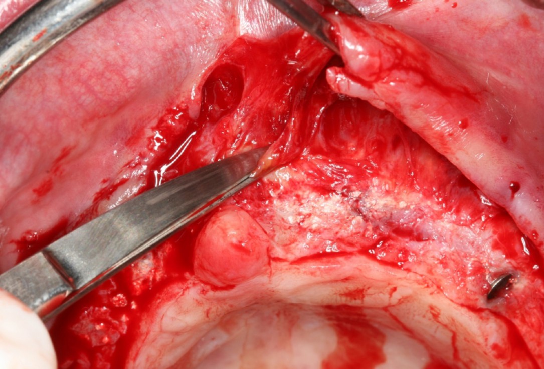
02/10 - Deep bone splitting with oscillating saw in regio 15 to 25Circular bone splitting with maxresorb® & collprotect® membrane - PD Dr. J. Neugebauer
-
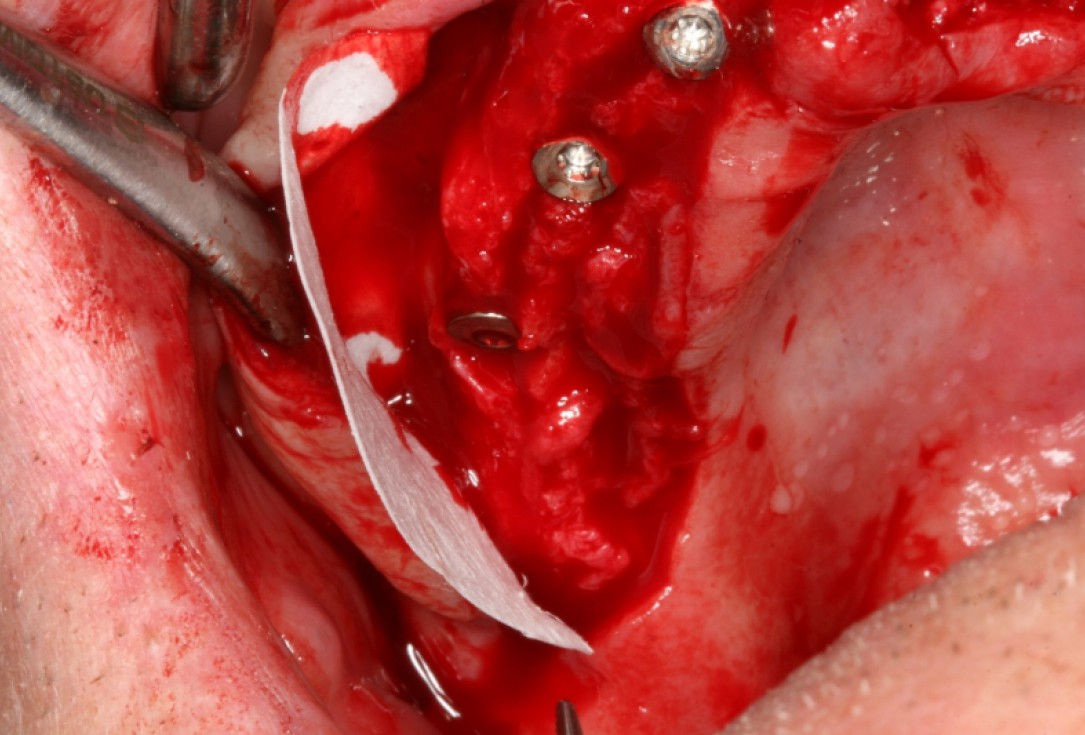
03/10 - Positioning of collprotect® membrane for application of bone graft materialCircular bone splitting with maxresorb® & collprotect® membrane - PD Dr. J. Neugebauer
-
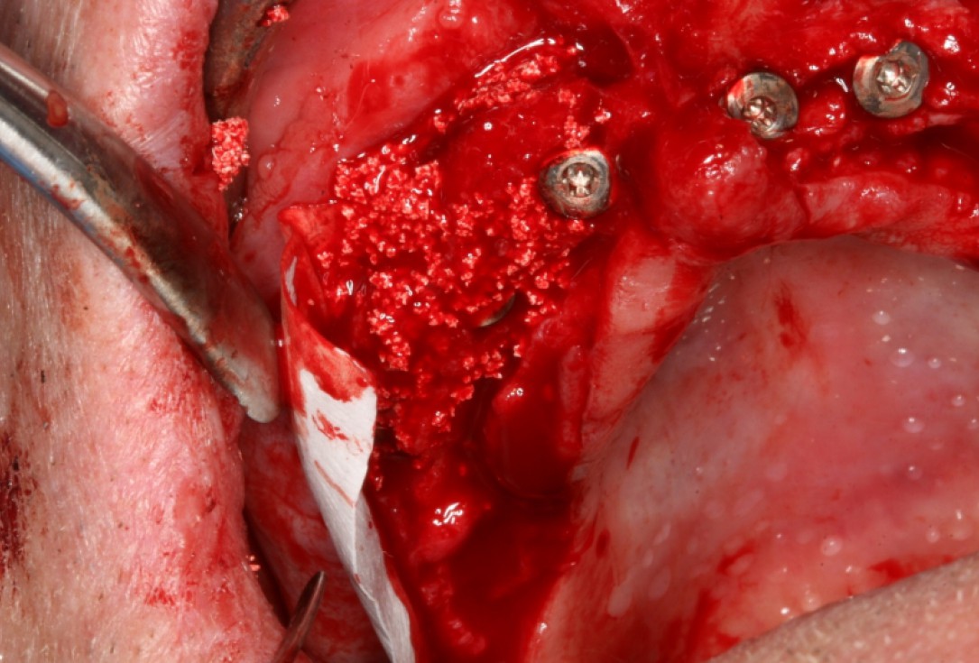
04/10 - Lateral deposition of maxresorb® to prevent resorption of the vestibular wallCircular bone splitting with maxresorb® & collprotect® membrane - PD Dr. J. Neugebauer
-
05/10 - Covering of the augmentation site with the initially inserted membraneCircular bone splitting with maxresorb® & collprotect® membrane - PD Dr. J. Neugebauer
-
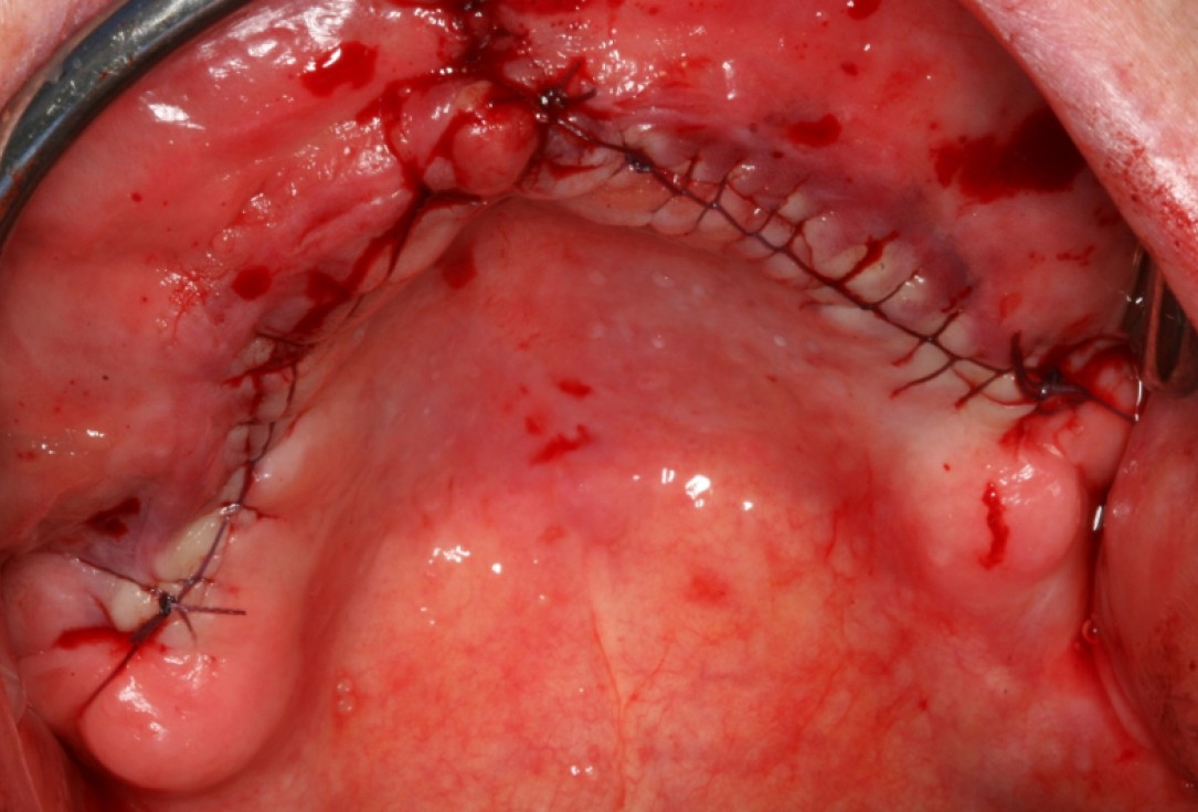
06/10 - Tight wound closure with a continuous seam following the periost splittingCircular bone splitting with maxresorb® & collprotect® membrane - PD Dr. J. Neugebauer
-
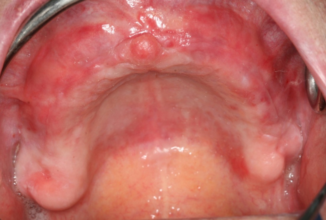
07/10 - Complication-free healing of the augmented ridgeCircular bone splitting with maxresorb® & collprotect® membrane - PD Dr. J. Neugebauer
-
08/10 - Re-entry surgery in combination with vestibuloplasty to form the vestibulumCircular bone splitting with maxresorb® & collprotect® membrane - PD Dr. J. Neugebauer
-
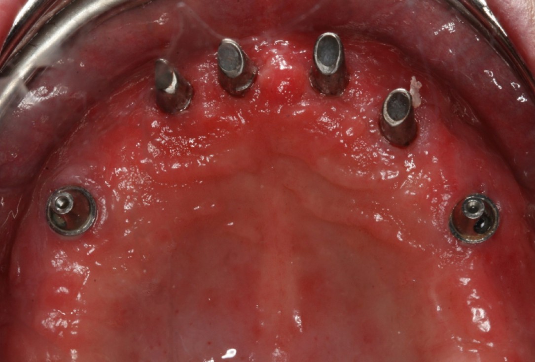
09/10 - Soft tissue situation after healing with inserted abutmentsCircular bone splitting with maxresorb® & collprotect® membrane - PD Dr. J. Neugebauer
-
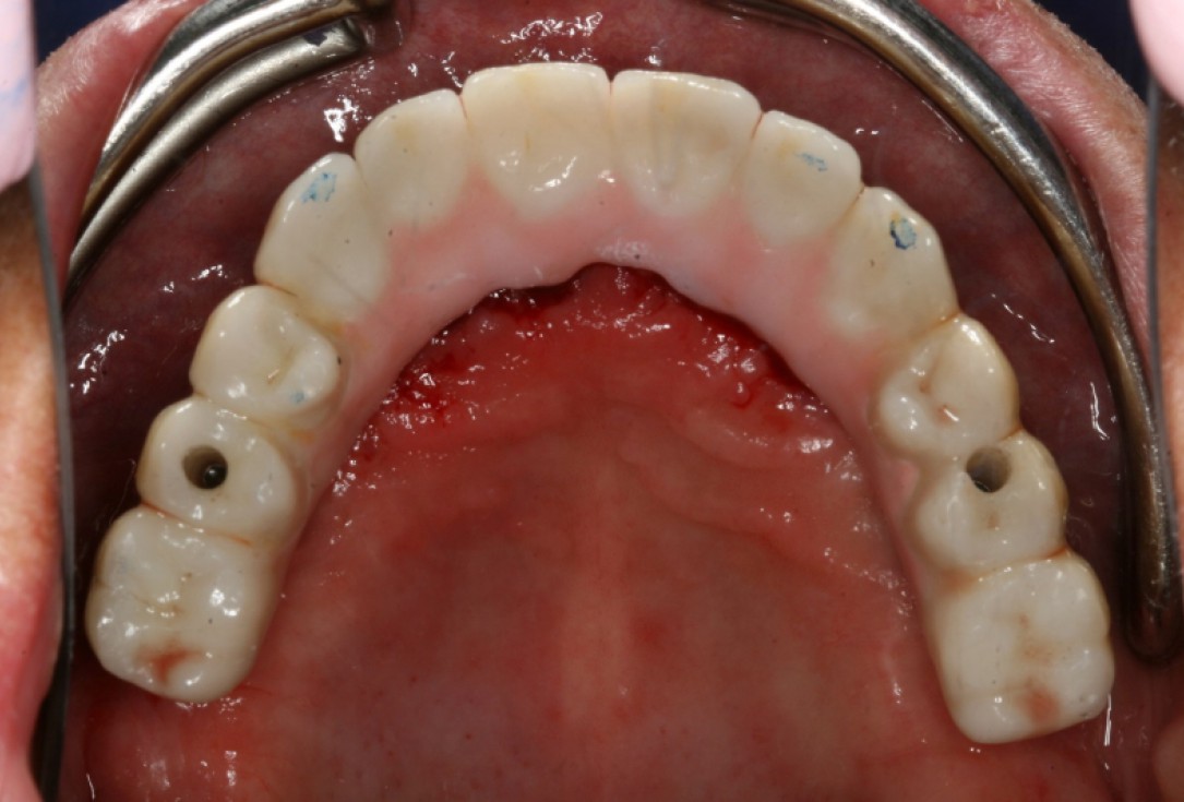
10/10 - Inserted bridge with terminally screwed and anteriorly cemented implantsCircular bone splitting with maxresorb® & collprotect® membrane - PD Dr. J. Neugebauer

Initial Orthopantomograph X-Ray

Pre-operative x-ray

X-ray control before tooth extraction

DVT image demonstrating horizontal and vertical amount of bone available

Surgical presentation of the alveolar ridge with reduced amount of horizontal bone available

DVT control after sinusitis surgery, residual bone height 1 mm

Clinical situation before extraction

DVT control after sinusitis surgery, residual bone height 1 mm

DVT image showing the reduced amount of bone available in the area of the mental foramen

X-ray shows a 3-dimensional periondontal defect

Initial situation: Inflammated tooth #12
