Vertical bone augmentation and broadening of attached gingiva using cerabone®, permamem® and mucoderm® - Dr. R. Naimoli
-
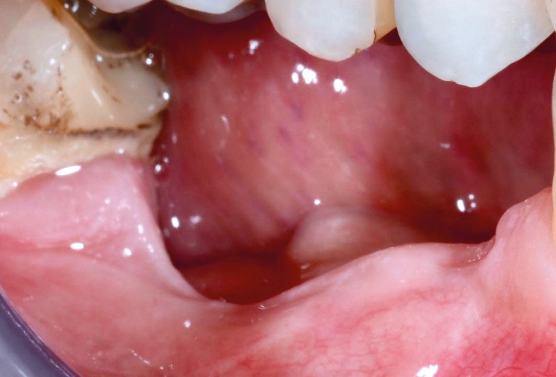
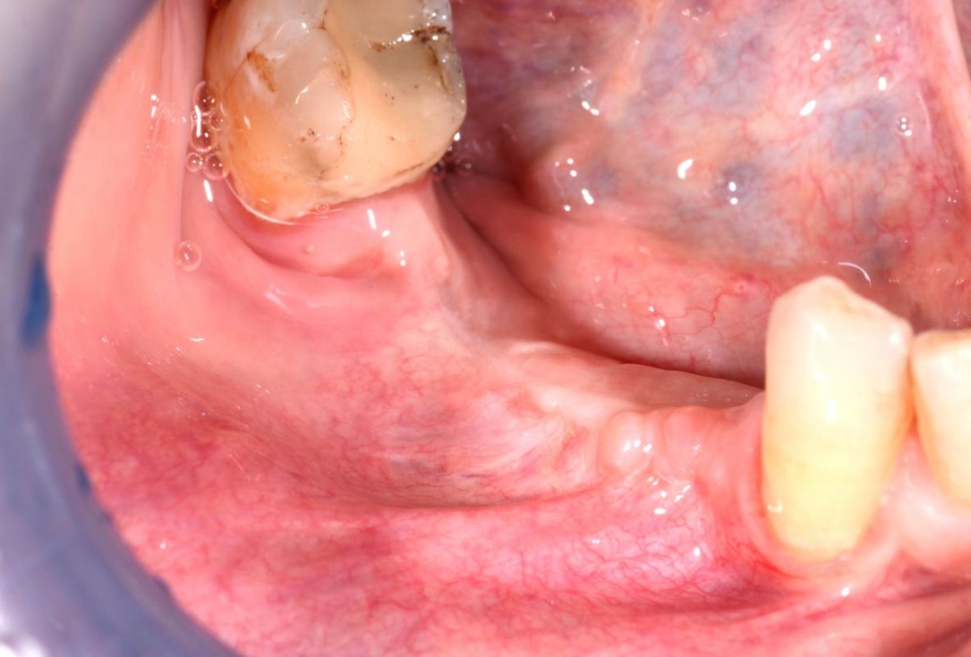
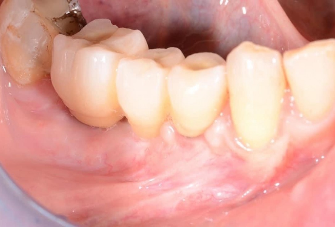
01/29 - Initial clinical situation with pronounced vertical and horizontal bone defectVertical bone augmentation and broadening of attached gingiva using cerabone®, permamem® and mucoderm® - Dr. R. Naimoli
-
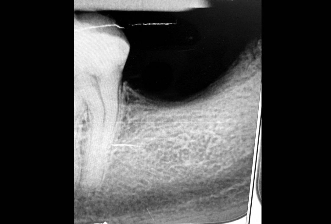
02/29 - Based on the intraoral x-ray a vertical bone defect of about 4 mm extending from 4.6 to 4.4 was determinedVertical bone augmentation and broadening of attached gingiva using cerabone®, permamem® and mucoderm® - Dr. R. Naimoli
-
03/29 - Preparation of a full-thickness flapVertical bone augmentation and broadening of attached gingiva using cerabone®, permamem® and mucoderm® - Dr. R. Naimoli
-
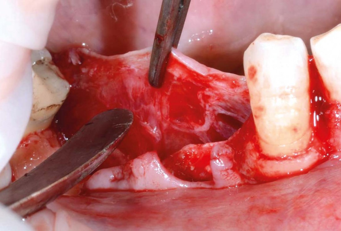
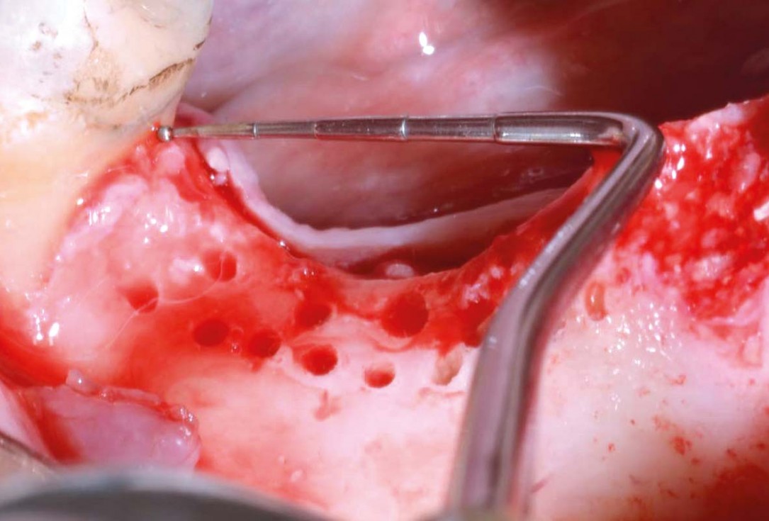
04/29 - Clinical view after exposure of the defect, to promote healing the local bone was perforatedVertical bone augmentation and broadening of attached gingiva using cerabone®, permamem® and mucoderm® - Dr. R. Naimoli
-
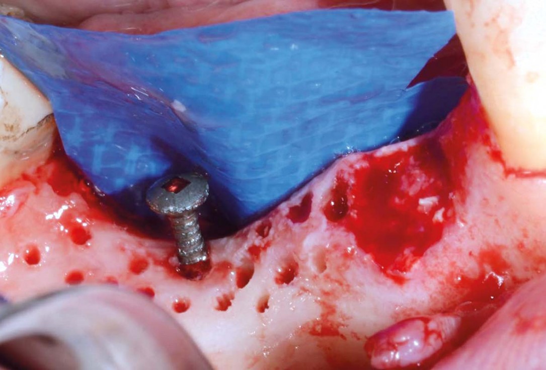
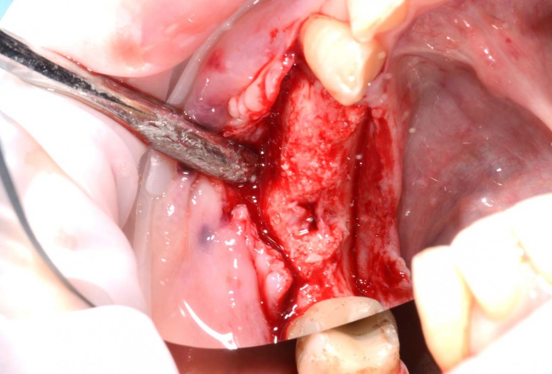
05/29 - Fixation of an osteosynthesis screw at the lowest point of the defect, lingual positioning of permamem®Vertical bone augmentation and broadening of attached gingiva using cerabone®, permamem® and mucoderm® - Dr. R. Naimoli
-
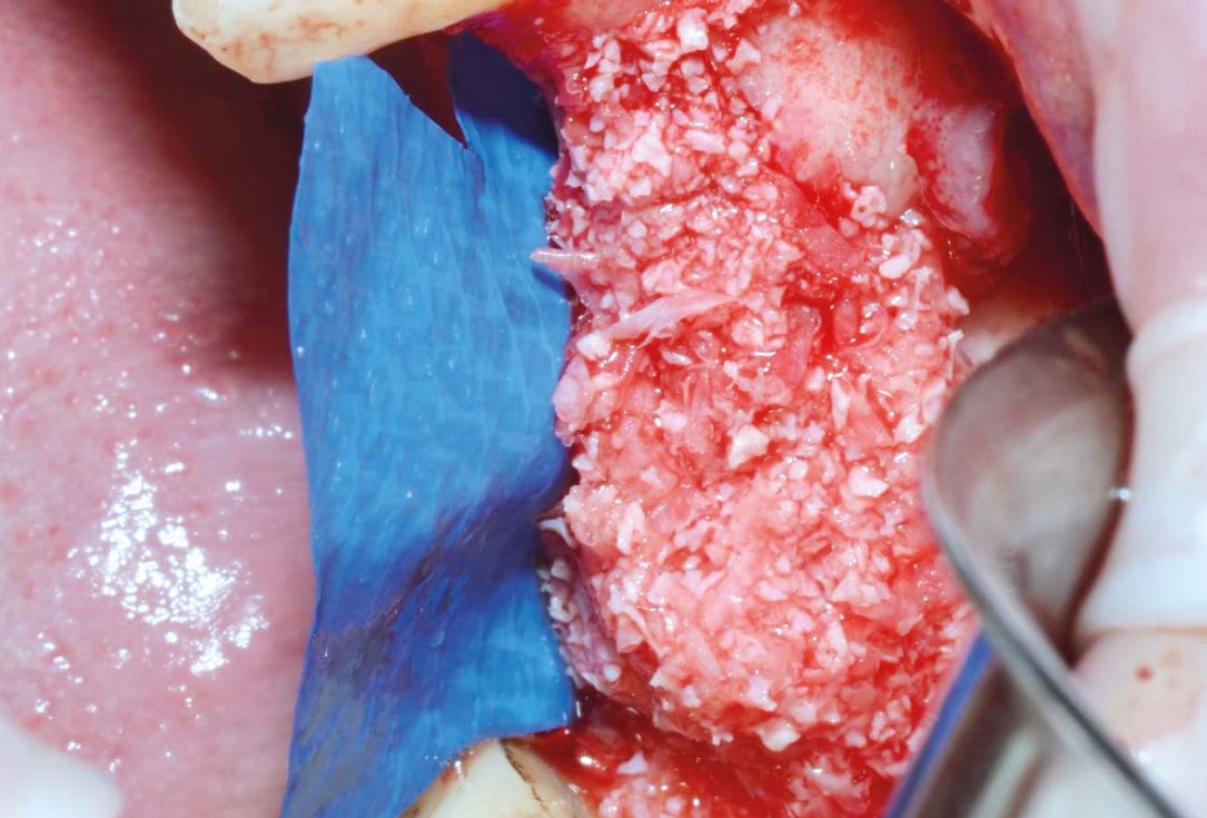
06/29 - Augmentation with cerabone® and autologous bone (ratio of 1:1)Vertical bone augmentation and broadening of attached gingiva using cerabone®, permamem® and mucoderm® - Dr. R. Naimoli
-
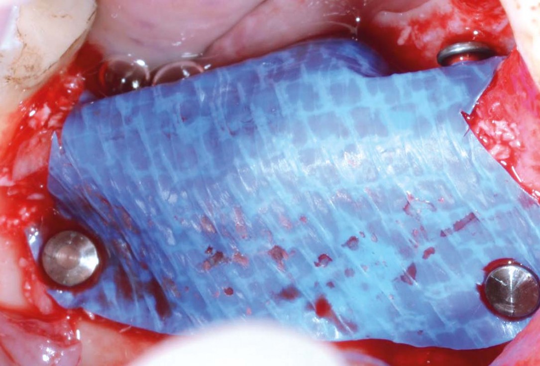
07/29 - Covering of the augmented area and buccal fixation of permamem® with titanium pinsVertical bone augmentation and broadening of attached gingiva using cerabone®, permamem® and mucoderm® - Dr. R. Naimoli
-
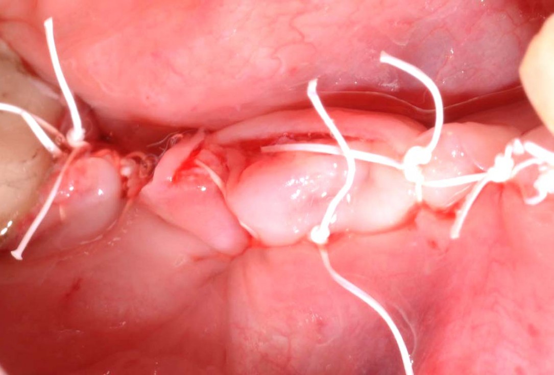
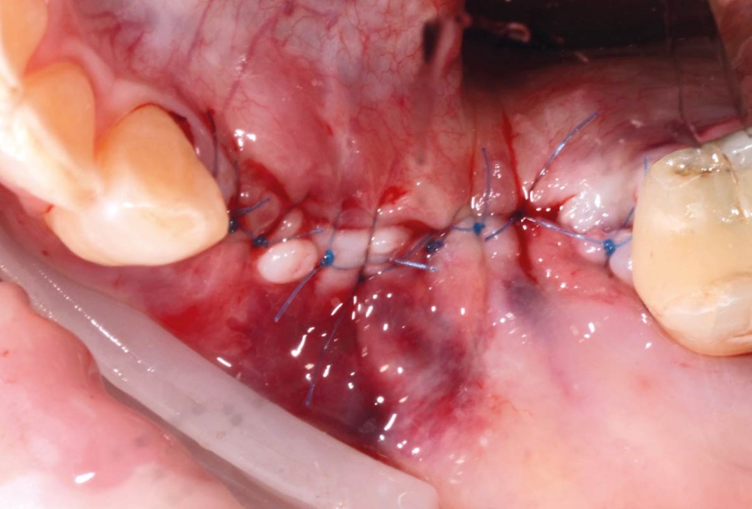
08/29 - Defect closure with non-absorbable PTFE suturesVertical bone augmentation and broadening of attached gingiva using cerabone®, permamem® and mucoderm® - Dr. R. Naimoli
-
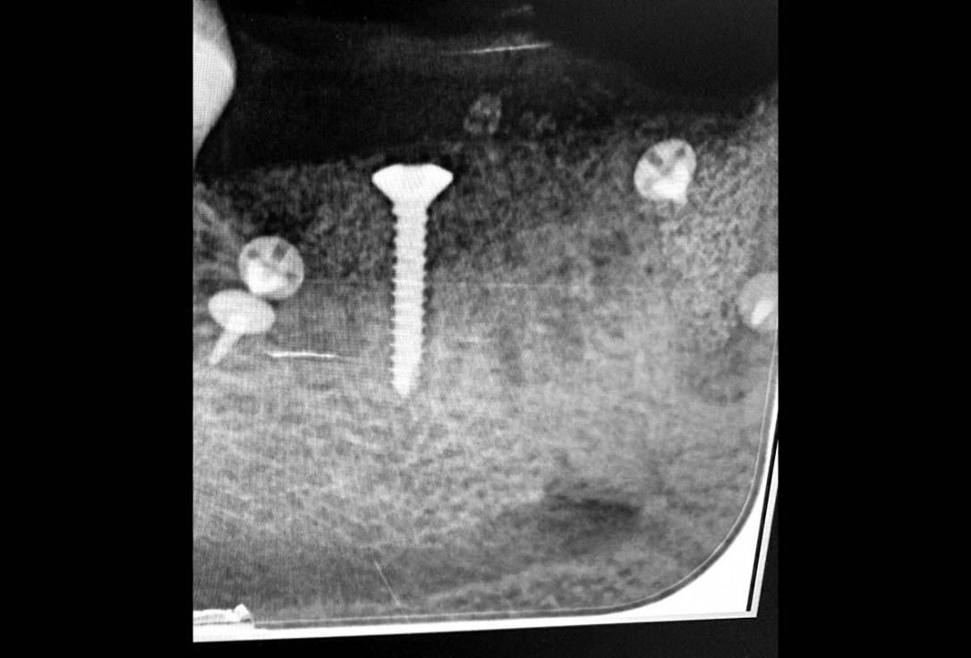
09/29 - Post-operative x-ray controlVertical bone augmentation and broadening of attached gingiva using cerabone®, permamem® and mucoderm® - Dr. R. Naimoli
-
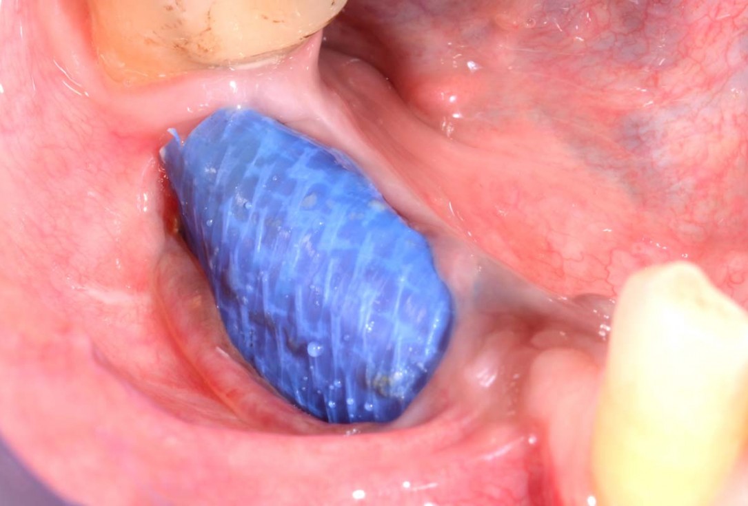
10/29 - Dehiscence of permamem® 3 months post-operatively. No signs of infection, the patient did not report painVertical bone augmentation and broadening of attached gingiva using cerabone®, permamem® and mucoderm® - Dr. R. Naimoli
-
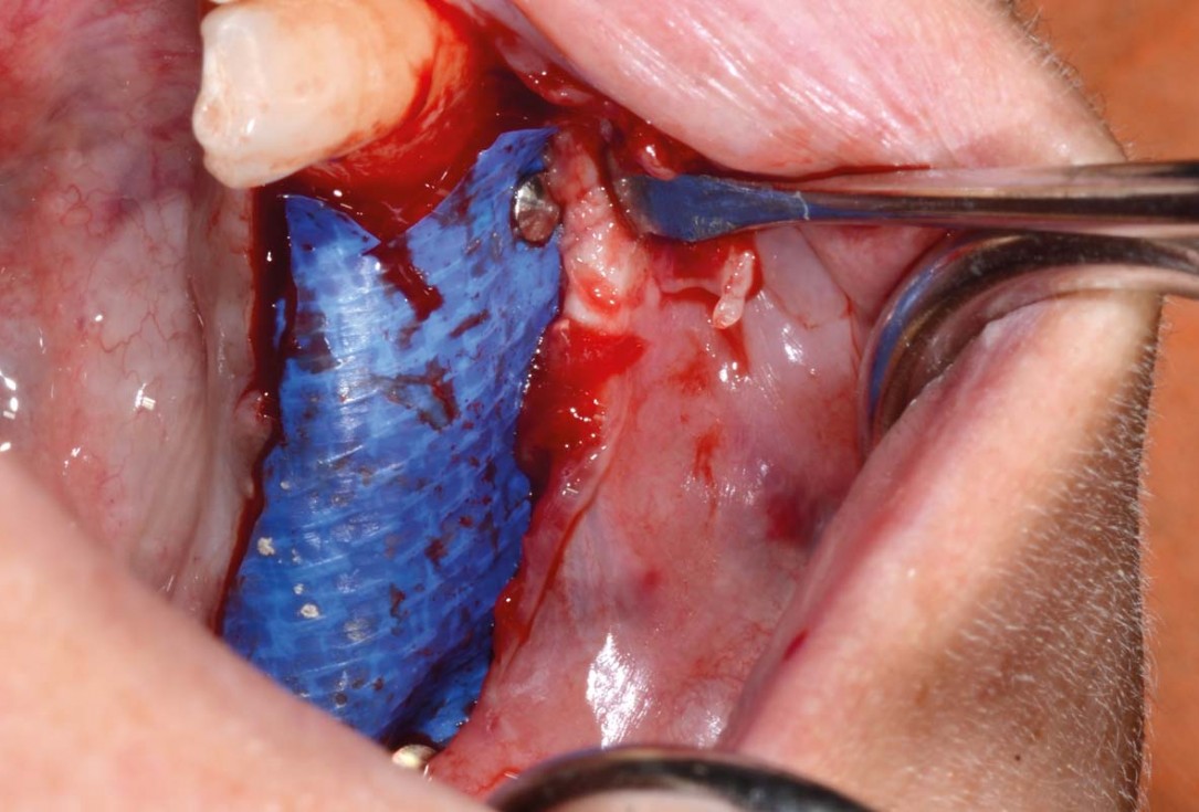
11/29 - Flap opening for removal of permamem®Vertical bone augmentation and broadening of attached gingiva using cerabone®, permamem® and mucoderm® - Dr. R. Naimoli
-
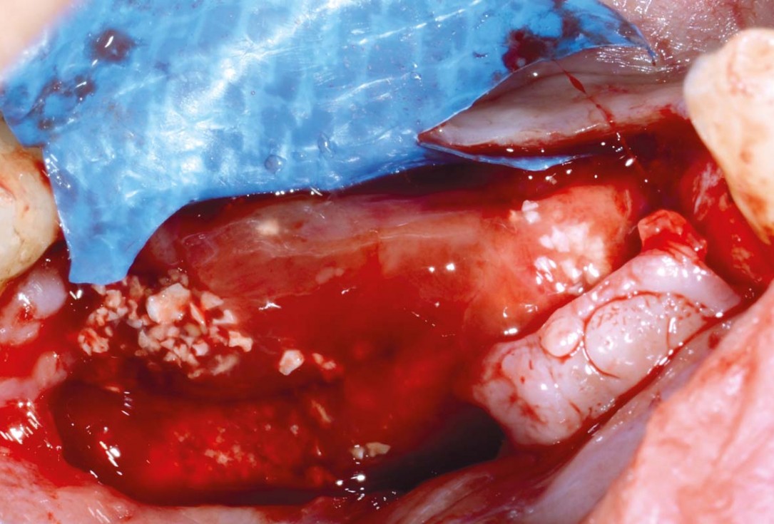
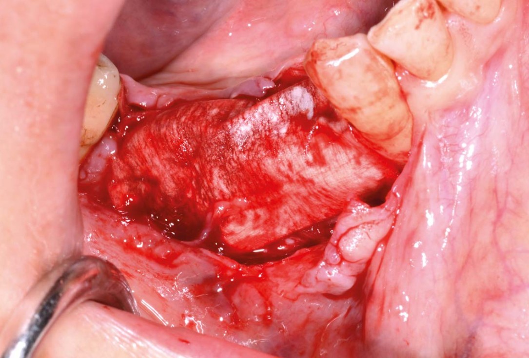
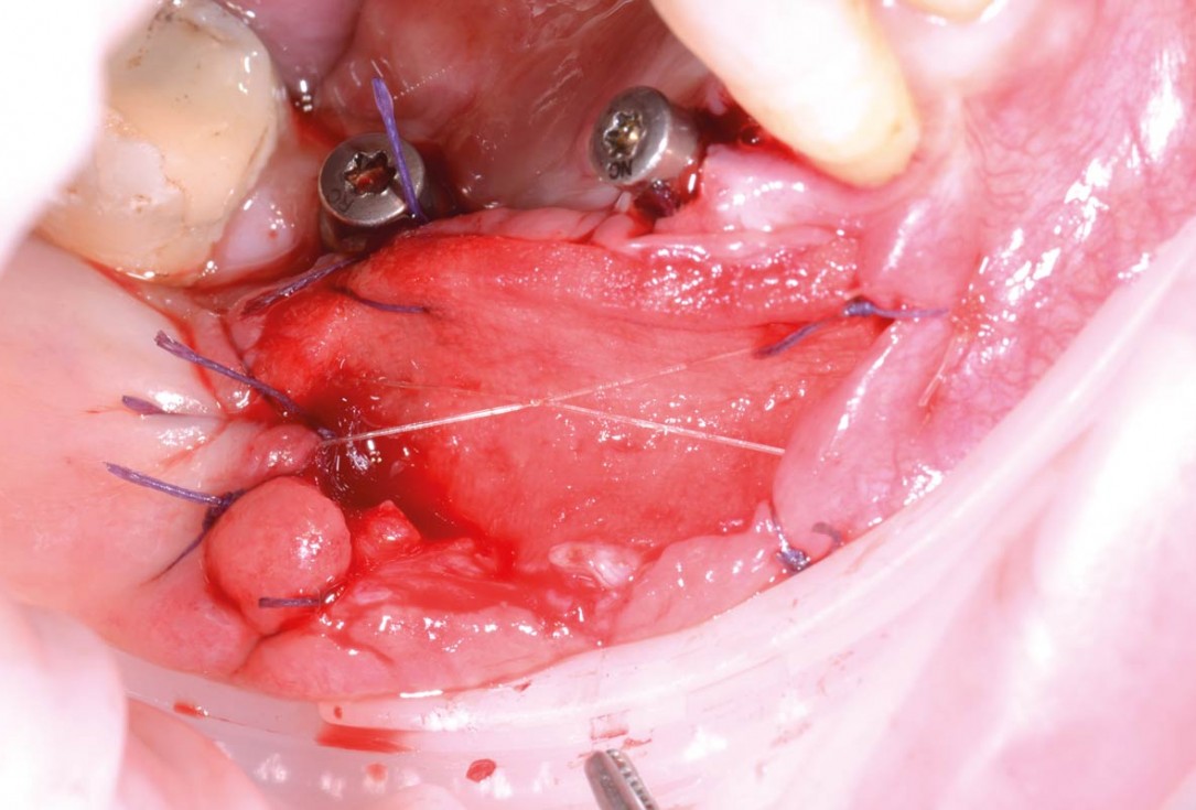
12/29 - Augmented area in maturation phase exposed under permamem®Vertical bone augmentation and broadening of attached gingiva using cerabone®, permamem® and mucoderm® - Dr. R. Naimoli
-
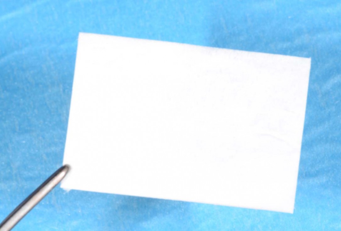
13/29 - Jason® membrane before hydrationVertical bone augmentation and broadening of attached gingiva using cerabone®, permamem® and mucoderm® - Dr. R. Naimoli
-
14/29 - Jason® membrane covering the augmented areaVertical bone augmentation and broadening of attached gingiva using cerabone®, permamem® and mucoderm® - Dr. R. Naimoli
-
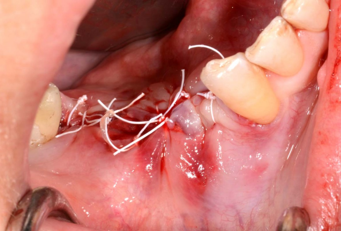
15/29 - Flap closureVertical bone augmentation and broadening of attached gingiva using cerabone®, permamem® and mucoderm® - Dr. R. Naimoli
-
16/29 - Clinical view at re-entry, 8 months post-opVertical bone augmentation and broadening of attached gingiva using cerabone®, permamem® and mucoderm® - Dr. R. Naimoli
-
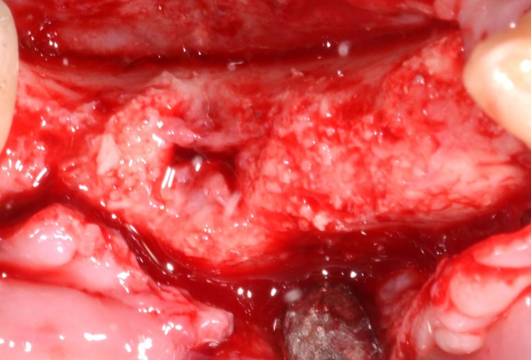
17/29 - Slight loss of the graft in regio 4.6, but still favorable for the insertion of the implantsVertical bone augmentation and broadening of attached gingiva using cerabone®, permamem® and mucoderm® - Dr. R. Naimoli
-
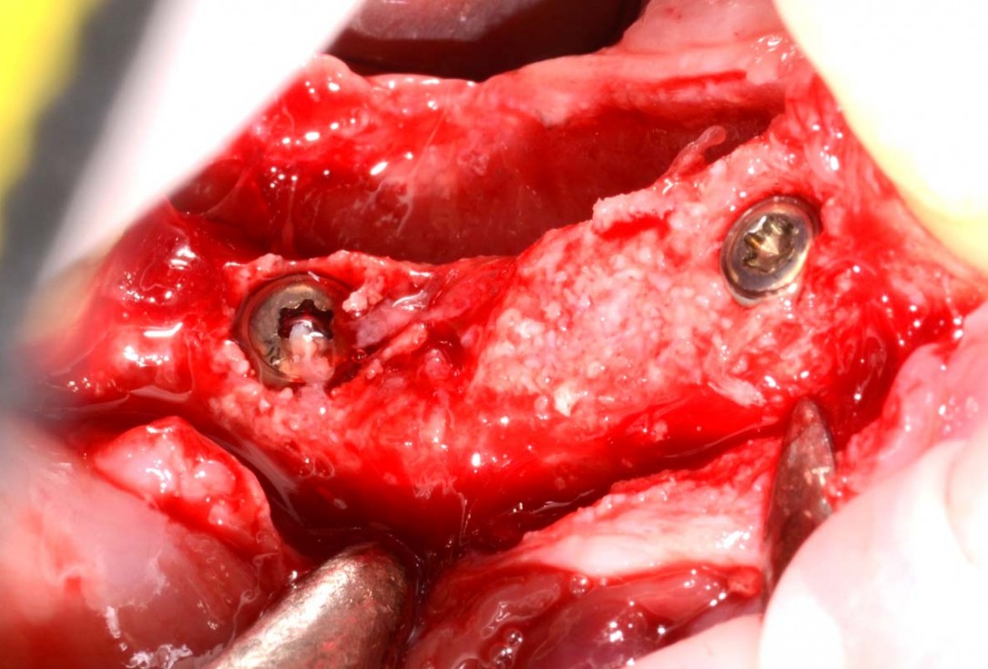
18/29 - Insertion of 2 Straumann BLT implants (regio 4.4: 3.3 x 10 mm, regio 4.6: 4.1x 8 mm), excellent primary stabilityVertical bone augmentation and broadening of attached gingiva using cerabone®, permamem® and mucoderm® - Dr. R. Naimoli
-
19/29 - Flap closureVertical bone augmentation and broadening of attached gingiva using cerabone®, permamem® and mucoderm® - Dr. R. Naimoli
-
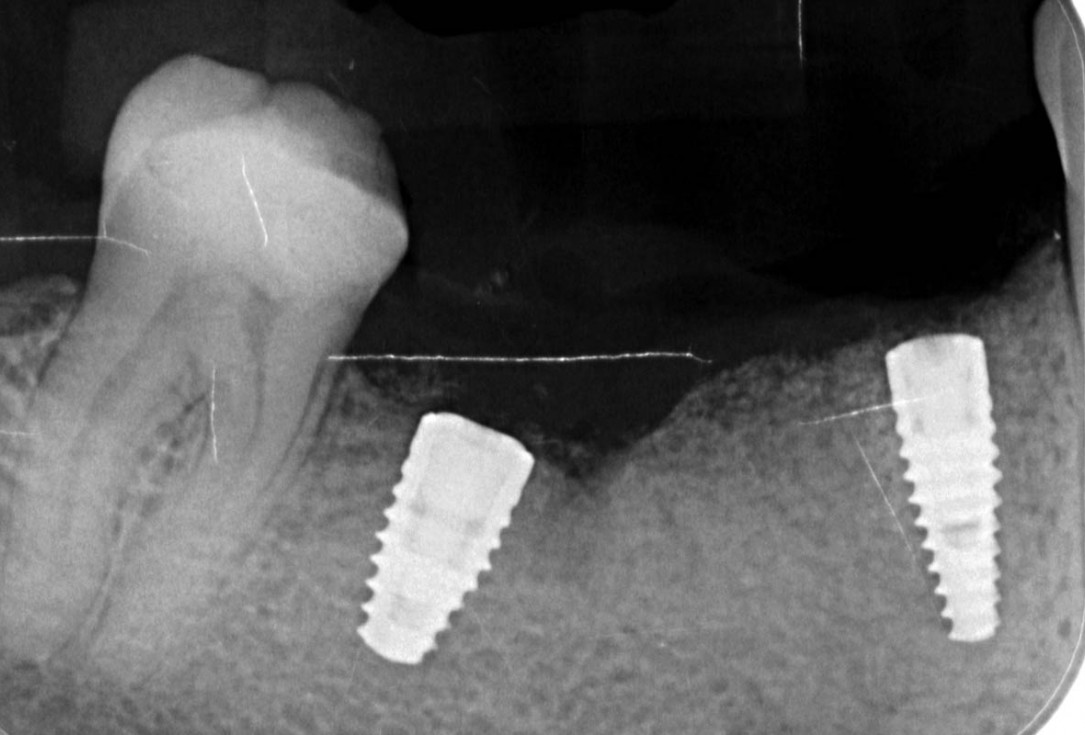
20/29 - X-ray controlVertical bone augmentation and broadening of attached gingiva using cerabone®, permamem® and mucoderm® - Dr. R. Naimoli
-
21/29 - Clinical situation 3 months after implantationVertical bone augmentation and broadening of attached gingiva using cerabone®, permamem® and mucoderm® - Dr. R. Naimoli
-
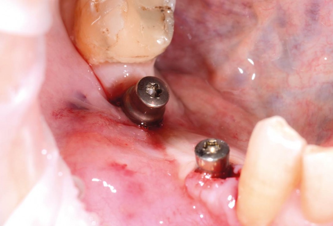
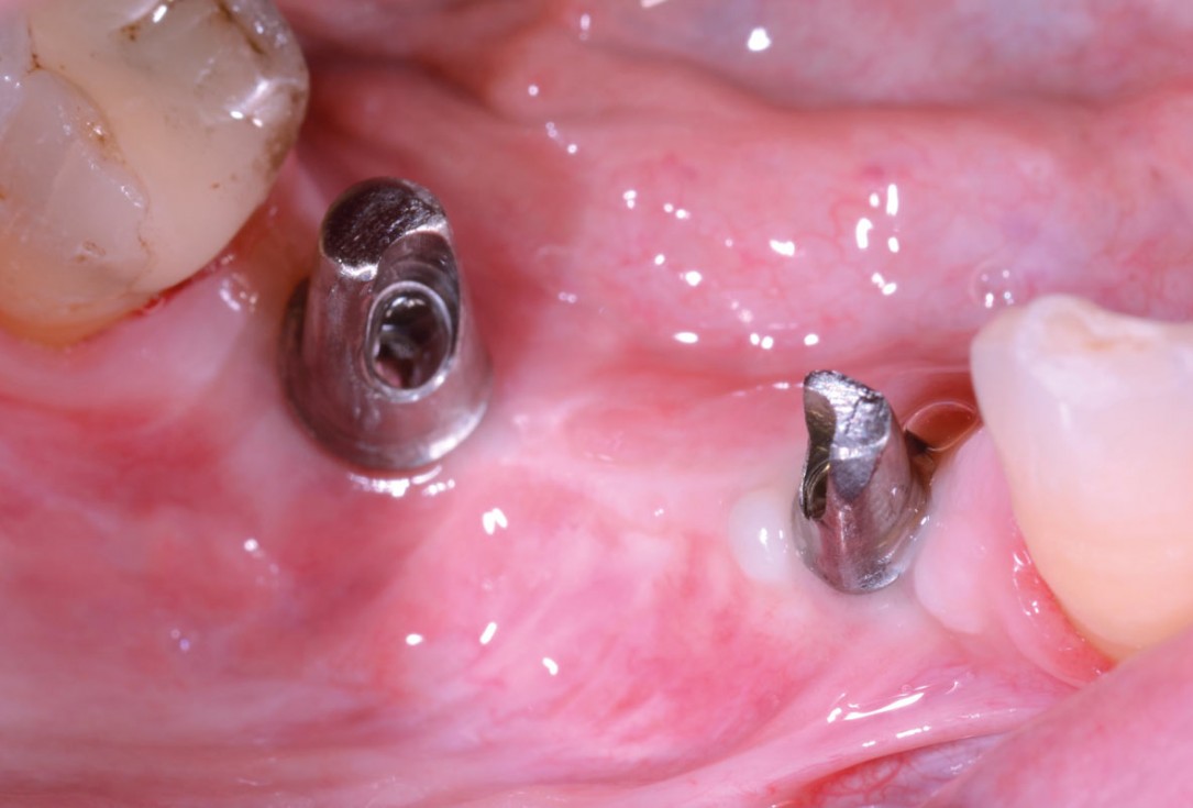
22/29 - Clinical view after insertion of the healing abutmentVertical bone augmentation and broadening of attached gingiva using cerabone®, permamem® and mucoderm® - Dr. R. Naimoli
-
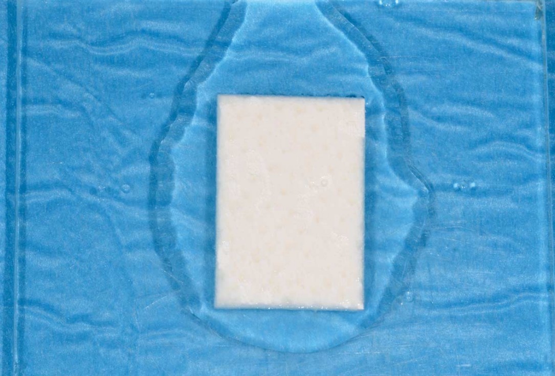
23/29 - Hydration of mucoderm®Vertical bone augmentation and broadening of attached gingiva using cerabone®, permamem® and mucoderm® - Dr. R. Naimoli
-
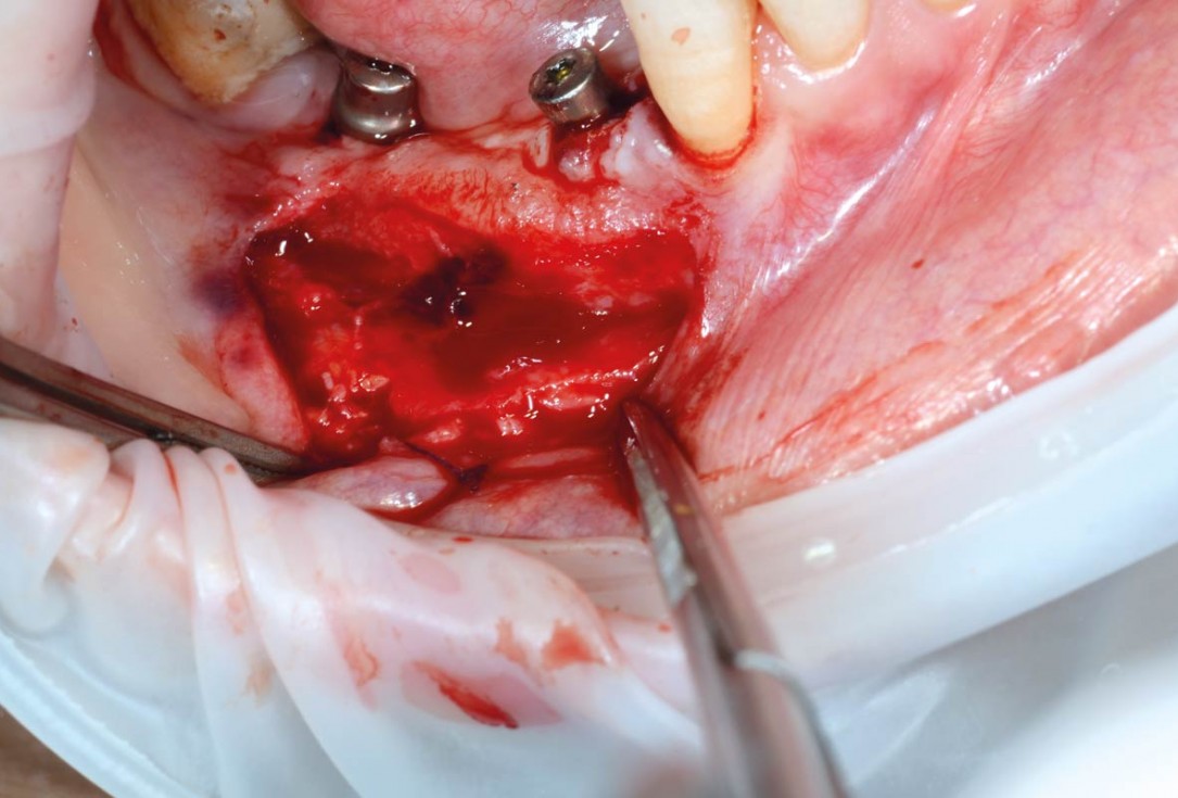
24/29 - Preparation of vestibular partial thickness flap (highlighting and isolation of mental nerve)Vertical bone augmentation and broadening of attached gingiva using cerabone®, permamem® and mucoderm® - Dr. R. Naimoli
-
25/29 - Fixation of mucoderm® to periosteum and flapVertical bone augmentation and broadening of attached gingiva using cerabone®, permamem® and mucoderm® - Dr. R. Naimoli
-
26/29 - Clinical view with broadened attached gingiva after 3 weeks, impression takingVertical bone augmentation and broadening of attached gingiva using cerabone®, permamem® and mucoderm® - Dr. R. Naimoli
-
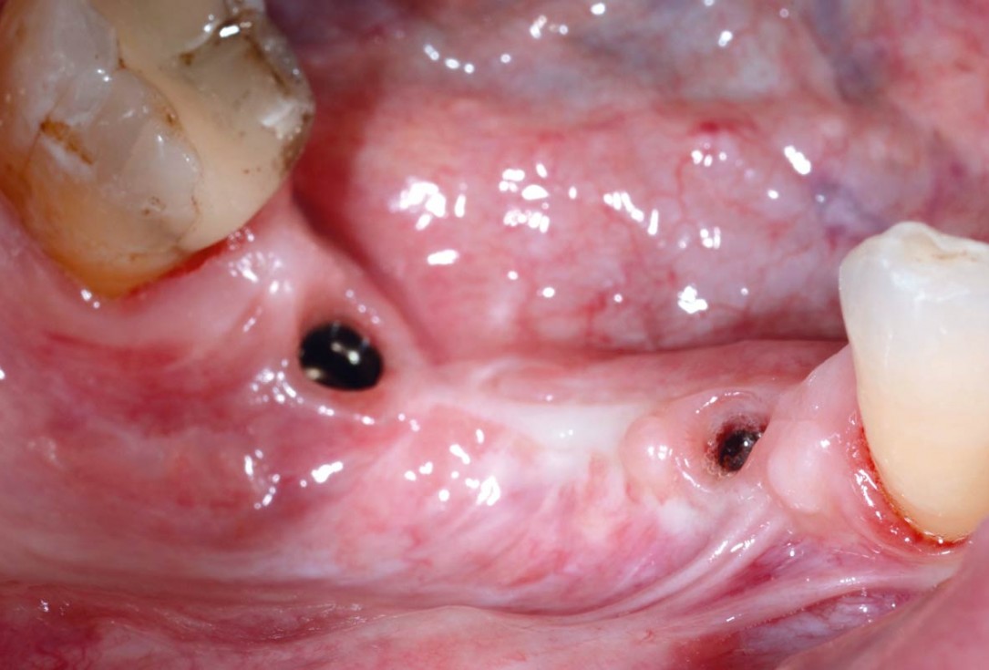
27/29 - Clinical view before installation of the fixed prosthetics, good amount of keratinized tissue around the implantsVertical bone augmentation and broadening of attached gingiva using cerabone®, permamem® and mucoderm® - Dr. R. Naimoli
-
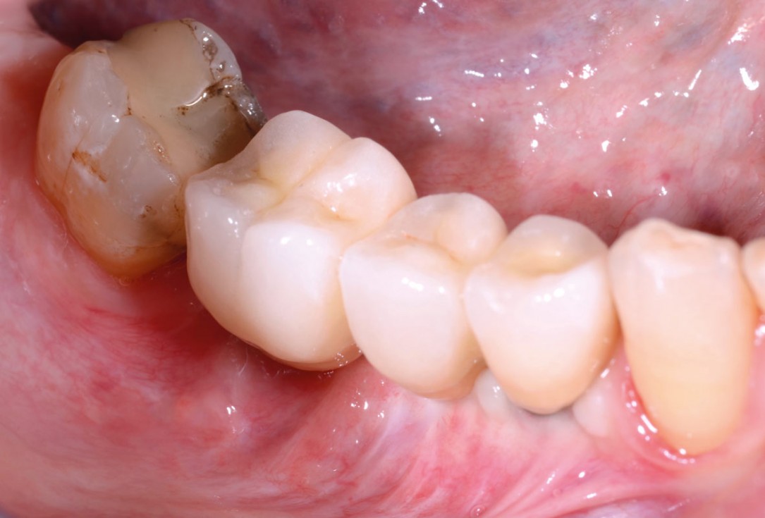
28/29 - Final prosthetic restorationVertical bone augmentation and broadening of attached gingiva using cerabone®, permamem® and mucoderm® - Dr. R. Naimoli
-
29/29 - Clinical view at control 3 month after final restorationVertical bone augmentation and broadening of attached gingiva using cerabone®, permamem® and mucoderm® - Dr. R. Naimoli

Initial clinical situation. Atrophic maxillary ridge.

Initial x-ray showing bone loss around implants placed 5 years ago in another dental clinic

Initial view of the case. Discoloration of 1.1 and mild class I gingival recession

Situation after tooth removal.

Initial clinical situation with gum recession and labial bone loss eight weeks following tooth extraction

Three implants placed in a narrow posterior mandible

Pre-operative clinical situation.

Clinical situation with narrow alveolar ridge in the lower jaw

Initial clinical situation showing bone wall defect.

Initial clinical situation.

Pre-surgical situation.

Initial situation: missing teeth #11 & 12 and badly broken #21 root

Pre-operative OPG shows deep vertical intrabony defects on the distal aspects of teeth 13 and 14.

Instable bridge situation with abscess formation at tooth #15 after apicoectomy

Initial clinical situation.

Implant insertion in atrophic alveolar ridge

Preoperative clinical situation

Pre-operative OPG

Pre-operative X-ray. Hopless tooth 21.

Pre-surgical situation. Teeth 26 and 27 missing.

Extraction of tooth 21 after endodontic treatment

Pre-surgical probing reveals a deep intrabony defect on the distal aspect of the upper canine.

Initial clinical situation with single tooth gap in regio 21

Pre-operative radiographic view. Intrabony defect on the distal aspect of the lateral incisor.

Clinical situation before extraction and implantation

Pre-operative radiographic view.
