Two-wall intrabony defect treated using cerabone® and Straumann® Emdogain® - Dr. D. Rakasevic & Prof. Dr. S. Jankovic
-
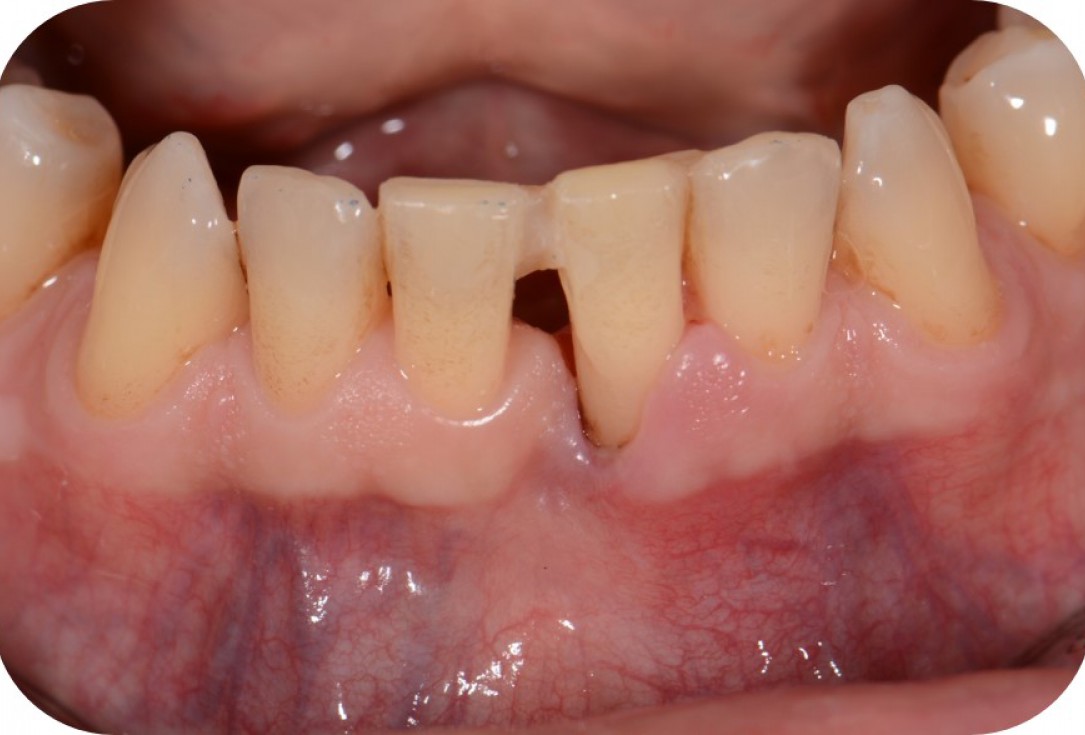
01/12 - Pre-operative clinical situation.Two-wall intrabony defect treated using cerabone® and Straumann® Emdogain® - Dr. D. Rakasevic & Prof. Dr. S. Jankovic
-
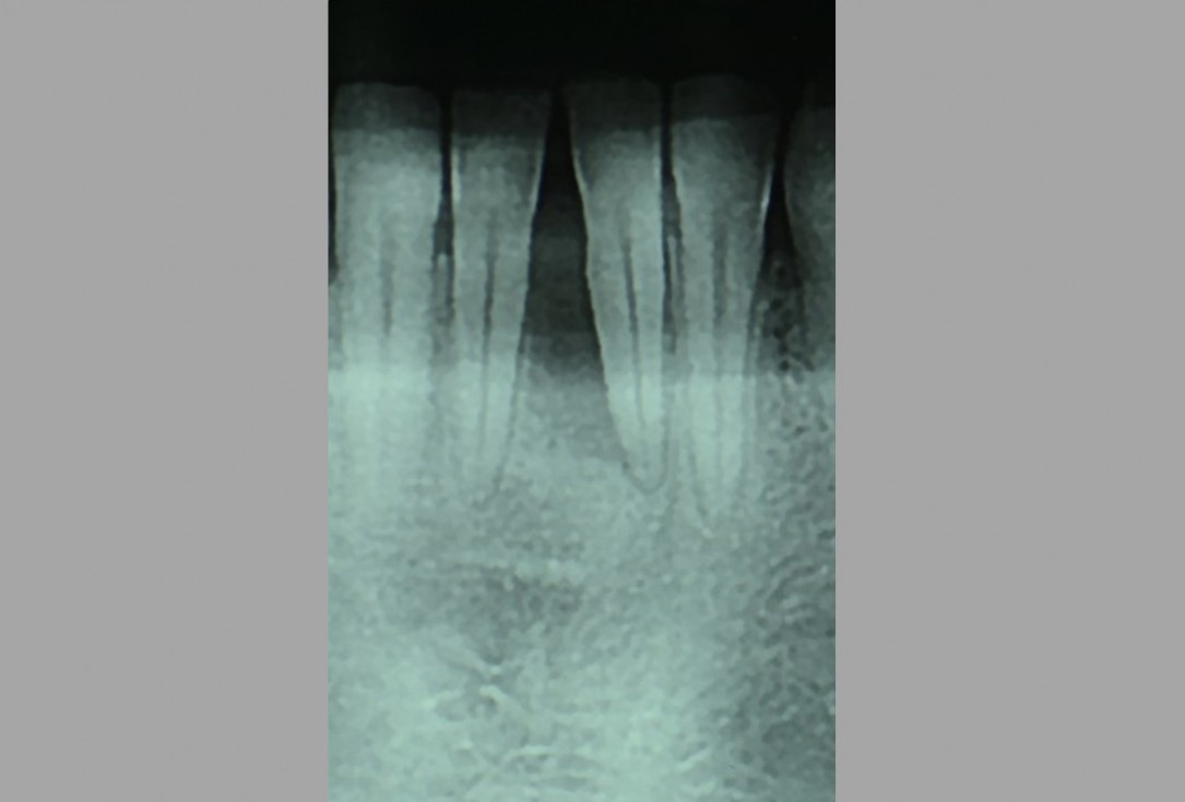
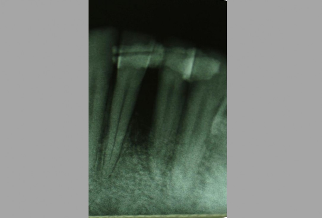
02/12 - Pre-operative radiograph. Deep intrabony defect at tooth 31.Two-wall intrabony defect treated using cerabone® and Straumann® Emdogain® - Dr. D. Rakasevic & Prof. Dr. S. Jankovic
-
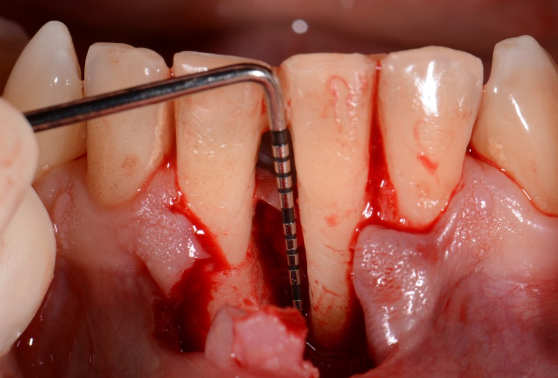
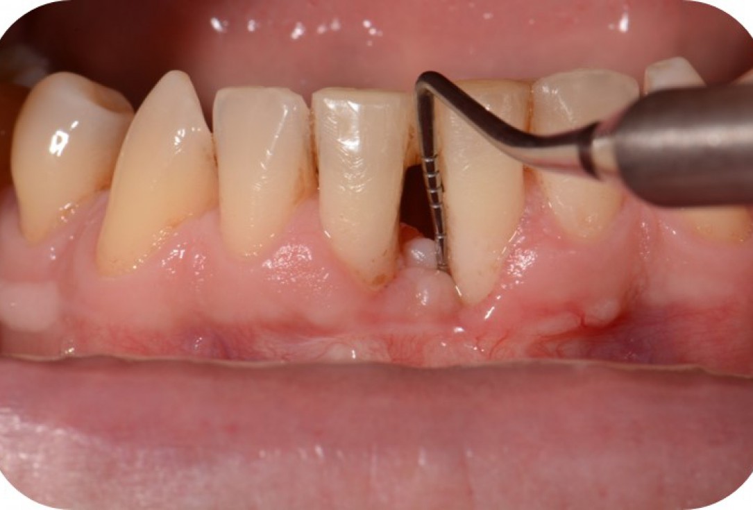
03/12 - Intra-operative view shows a deep two-wall intrabony defect mesial to tooth 31 (PPD 13 mm).Two-wall intrabony defect treated using cerabone® and Straumann® Emdogain® - Dr. D. Rakasevic & Prof. Dr. S. Jankovic
-
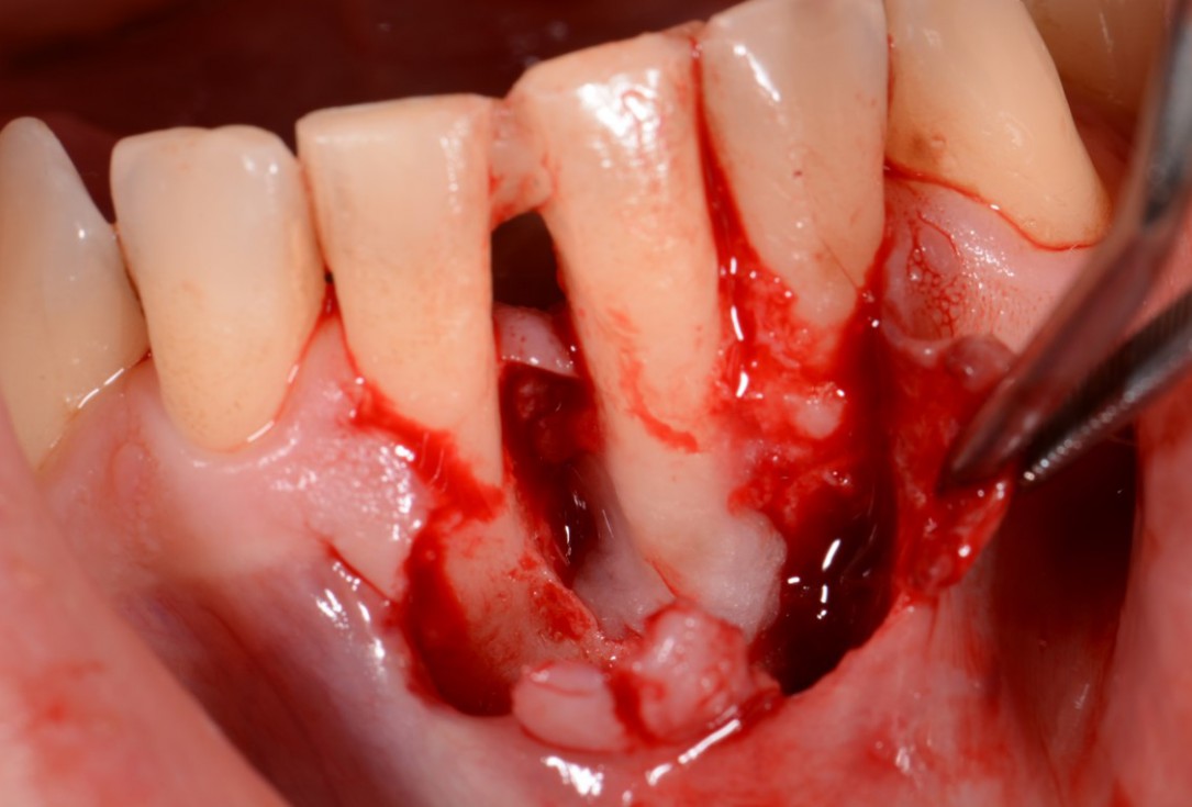
04/12 - Application of Straumann® Emdogain® to the exposed root surface.Two-wall intrabony defect treated using cerabone® and Straumann® Emdogain® - Dr. D. Rakasevic & Prof. Dr. S. Jankovic
-
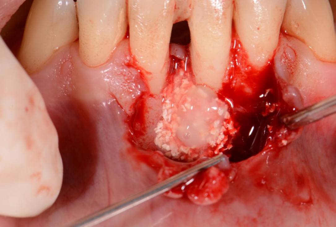
05/12 - Application of small cerabone® granules mixed with Straumann® Emdogain®.Two-wall intrabony defect treated using cerabone® and Straumann® Emdogain® - Dr. D. Rakasevic & Prof. Dr. S. Jankovic
-
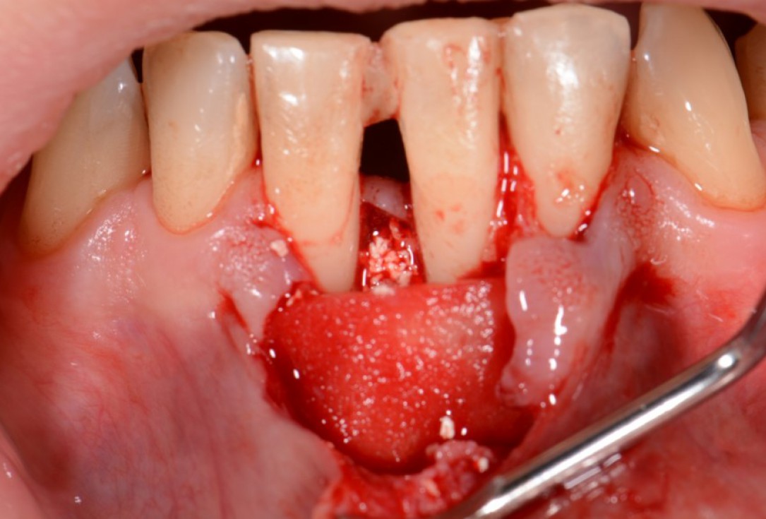
06/12 - Soft tissue augmentation with mucoderm® collagen matrix.Two-wall intrabony defect treated using cerabone® and Straumann® Emdogain® - Dr. D. Rakasevic & Prof. Dr. S. Jankovic
-
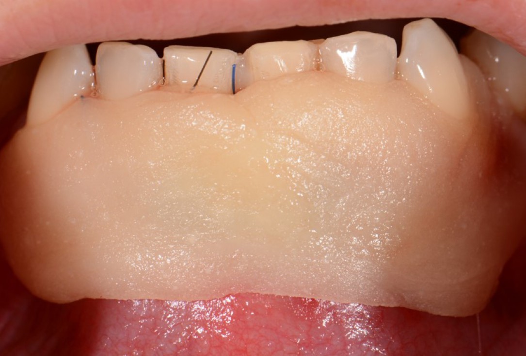
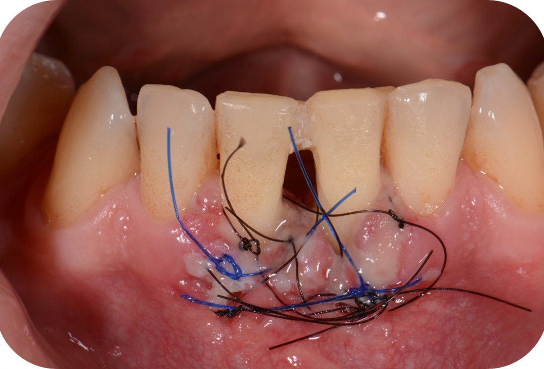
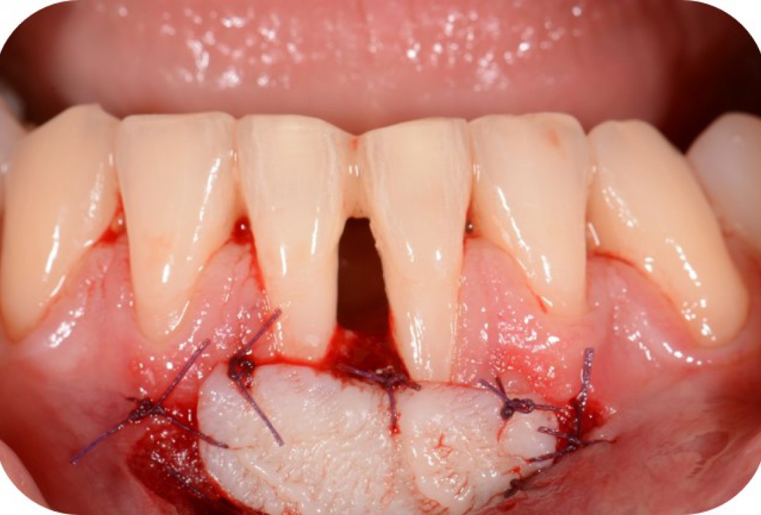
07/12 - Primary wound closure and application of a periodontal dressing.Two-wall intrabony defect treated using cerabone® and Straumann® Emdogain® - Dr. D. Rakasevic & Prof. Dr. S. Jankovic
-
08/12 - Clinical situation 1 month post-operative.Two-wall intrabony defect treated using cerabone® and Straumann® Emdogain® - Dr. D. Rakasevic & Prof. Dr. S. Jankovic
-
09/12 - Augmentation of keratinized gingiva with a free mucosal graft.Two-wall intrabony defect treated using cerabone® and Straumann® Emdogain® - Dr. D. Rakasevic & Prof. Dr. S. Jankovic
-
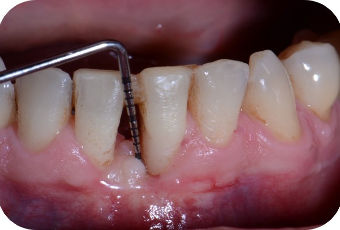
10/12 - Clinical situation 3 months post-operative with 5 mm probing pocket depth.Two-wall intrabony defect treated using cerabone® and Straumann® Emdogain® - Dr. D. Rakasevic & Prof. Dr. S. Jankovic
-
11/12 - Clinical situation 6 months post-operative.Two-wall intrabony defect treated using cerabone® and Straumann® Emdogain® - Dr. D. Rakasevic & Prof. Dr. S. Jankovic
-
12/12 - Radiograph shows osseous fill of the defect 6 months post-operative.Two-wall intrabony defect treated using cerabone® and Straumann® Emdogain® - Dr. D. Rakasevic & Prof. Dr. S. Jankovic

Initial clinical situation. Atrophic maxillary ridge.

Initial x-ray showing bone loss around implants placed 5 years ago in another dental clinic

Initial view of the case. Discoloration of 1.1 and mild class I gingival recession

Situation after tooth removal.

Initial clinical situation with gum recession and labial bone loss eight weeks following tooth extraction

Three implants placed in a narrow posterior mandible

Implant insertion in atrophic alveolar ridge

Preoperative clinical situation

Pre-operative OPG

Initial clinical situation.

Pre-surgical situation.

Initial situation: missing teeth #11 & 12 and badly broken #21 root

Pre-operative OPG shows deep vertical intrabony defects on the distal aspects of teeth 13 and 14.

Instable bridge situation with abscess formation at tooth #15 after apicoectomy

Initial clinical situation.

Pre-operative radiographic view. Intrabony defect on the distal aspect of the lateral incisor.

Clinical situation before extraction and implantation

Pre-operative radiographic view.

Situation after tooth extraction.

Pre-operative X-ray. Hopless tooth 21.

Pre-surgical situation. Teeth 26 and 27 missing.

Extraction of tooth 21 after endodontic treatment

Pre-surgical probing reveals a deep intrabony defect on the distal aspect of the upper canine.

Initial clinical situation with single tooth gap in regio 21

Clinical situation with narrow alveolar ridge in the lower jaw

Initial clinical situation showing bone wall defect.
