cerabone® - natural bovine bone grafting material

cerabone®

Material de injerto óseo bovino natural
Gracias a su fiabilidad y predecibilidad terapéutica, el material de elección para la mayoría de los dentistas es el injerto óseo de origen bovino. cerabone® es un injerto óseo altamente fiable, dimensionalmente estable y seguro. Es derivado de la fase mineral del hueso bovino, que presenta una gran similitud con el hueso humano en términos de composición química, porosidad y estructura superficial.
  • Preservación alveolar y de cresta
  • Afectaciones de furca (clases I-II)
  • Aumento de cresta
  • Defectos periimplantarios
  • Defectos intraóseos (1-3 paredes)
  • Elevación de seno

Rehidratación

La rehidratación de los gránulos en sangre del defecto o suero salino no es necesaria, pero simplifica tanto el manejo como su aplicación, dado que las partículas de cerabone® humectadas tienden a pegarse entre sí.

Compresión de las partículas

Durante la aplicación, evite comprimir excesivamente las partículas; unas partículas menos condensadas dejan más espacio para la penetración de vasos sanguíneos y la formación de nueva matriz ósea.

Tiempo de integración

Se recomienda un tiempo de integración de como mínimo 6 meses antes de la reentrada para asegurar una integración estable de las partículas.

Mezcla del material

Mezclar cerabone® con hueso autólogo aporta una actividad biológica (gracias a las propiedades osteoinductivas y osteogénicas del hueso autólogo) que contribuye a una regeneración más rápida y una mejor formación de hueso nuevo.

Regeneration of a 9 mm vertical bone defect with cerabone®, autologous bone and S-PRF –  Dr. A. Eslava

Initial x-ray showing bone loss around implants placed 5 years ago in another dental clinic

cerabone® and mucoderm® for immediacy in esthetic zone -Dr. M Motta

Initial view of the case. Discoloration of 1.1 and mild class I gingival recession

GBR with cerabone® and Jason® membrane in the front tooth region - Dr. H. Maghaireh

Initial clinical situation with gum recession and labial bone loss eight weeks following tooth extraction
botiss cerabone® & Jason® membrane for horizontal augmentation - Clinical case by Dr. M. Steigmann

Three implants placed in a narrow posterior mandible

botiss cerabone® & collprotect® membrane for GBR - Clinical case by Dr. V. Kalenchuk

Clinical situation with narrow alveolar ridge in the lower jaw

Sinus Floor Elevation with maxgraft® bonering and subcrestal implantation in an eggshell thin sinus - Dr. K. Chmielewski

Initial situation: X-ray scan reveals eggshell thin sinus floor (1-3 mm) on both sites of the maxilla; green areas indicate the planned maxgraft® bonerings and red areas the planned implants

Immediate implant placement using cerabone® and collagen fleece - Dr. D. Jelušić

Pre-operative situation showing tooth 21 with deep periodontal pocket. Tooth presented with mobility grade III.

botiss-cerabone-permamem-maxgraft-mucoderm-horizontal-gbr-maghaireh

Initial situation - A young female 34 years old lost her front teeth in an surfing accident and she had a 5 unit bridge supported by her upper left lateral and right canine. The restoration failed and both supporting crowns have exposed and leaking margins.

Reconstruction of Anterior Maxilla with maxgraft® bonebuilder – Dr. B. Han

Loss of teeth in anterior maxilla caused by periodontitis

GBR and soft tissue augmentation with cerabone® and mucoderm® - H. Maghaireh & V. Ivancheva

Initial situation: missing teeth #11 & 12 and badly broken #21 root

botiss cerabone® & Jason® membrane for GBR - Clinical case by Prof. Dr. Dr. D. Rothamel

Instable bridge situation with abscess formation at tooth #15 after apicoectomy

botiss cerabone® & Jason® membrane for GBR - Clinical case by Dr. S. Kovalevsky

Implant insertion in atrophic alveolar ridge

Advanced vertical augmentation in posterior maxilla with maxgraft® bonering - Dr. A. Isser

Initial situation 57-year old female patient. X-ray scan reveals severe bone loss due to inflammation in region 13. Treatment plan was extraction of teeth 13 and 14 and augmentation after healing.

Socket preservation with cerabone® - Dr. P. Kämmerer

Extraction socket grafted with cerabone.

Socket preservation using cerbaone® and permamem® - Dr. A. Caiazzo

Grafting of the extraction socket with small cerabone® granules.

GBR with Jason membrane® and cerabone® - D. Fontana

Lateral view of the defect in the posterior right maxilla.

Sinus lift one-stage with cerabone® and collprotect® membrane - Dr. V. Kalenchuk

Clinical situation of the edentulous distal maxilla before the surgery

botiss cerabone for ridge splitting and augmentation of an atrophic alveolar ridge - Clinical case by Dr. V. Moshirabadi

Situation before augmentation, atrophic alveolar ridge

Block augmentation with maxgraft® and cerabone® – Dr. F. Kloss

Initial CBCT scan - Fracture of left maxillary incisor and loss of buccal wall

Maxillary bone reconstruction in the anterior area with cerabone and Jason membrane - Dr. P.-Y. Gegout & Prof. O. Huck

Pre-operative: loss of interdental papilla between 12 and 11 associated with gingival inflammation and pus

Treatment of a maxillary cyst with cerabone® and Jason® membrane - Dr. D. Mari

Preoperative x-ray, multiple residual cysts of the upper jaw

Treatment of vertical bone loss by using cerabone®, autologous bone and Jason® membrane - Prof. Rafael Ortega Lopes

The patient presented with severe pain in the lateral incisor and a deficient adhesive provisional. Bruxism resulted in canine loss and premature contact in the lateral incisor.

Intrabony defect treated using collprotect® membrane & cerabone® (1) - Cosgarea & Sculean

Pre-surgical probing reveals a deep intrabony defect on the distal aspect of the upper canine.

botiss cerabone® & Jason® membrane for block augmentation with autologous bone blocks - clinical case by Dr. S. Stavar

Initial clinical situation with single tooth gap in regio 21

botiss cerabone® & collagen fleece for immediate implantation - Clinical case by Dr. D. Jelušić

Clinical situation before extraction and implantation

botiss cerabone® & Jason® membrane for GBR - clinical case by Dr. S. Stavar

Initial clinical situation with broken bridge abutment in regio 12 and tooth 21 not worth preserving

Restoration of all four incisors with two maxgraft® bonering - Dr. B Giesenhagen

Initial situation pre-op: Central incisors with mobility 3

Block augmentation with maxgraft® in the maxilla - Dr. R. Cutts

Initial situation: 40 year old female patient with extensive scar tissue after several surgeries restored with a Rochette bridge

Ridge augmentation with maxgraft® bonebuilder and sinus floor elevation – Dr. K.P. Schiechl

Initial clinical situation: Bone defect in the upper right maxilla (teeth #14-16)

Horizontal ridge augmentation with maxgraft® cortico - M.Sc. E. Kapogianni

OPG of the initial situation – provision of missing denture in regio 44 to 47 by a resin-retained bridge

Regenerative corticotomy to compensate lower incisor malocclusion with cerabone® and mucoderm®

Initial view of the clinical case: Class III malocclusion
Treatment plan: Regenerative corticotomy (PAOO)

Socket preservation with cerabone® - Dr. Rahib Adi Nader

Situation before extraction with single tooth crowns on 21 and 22

Immediate implant placement and correction of horizontal and vertical bone loss using an allograft bone ring, cerabone® and Jason® membrane - Drs. Miller and Korn

The patient presented with pathologic mobility of upper left central incisor. Radiographic examination revealed significant circumferential attachment loss with an unfavorable crown to root ratio.

Block grafting in the aesthetic zone with maxgraft®, Jason® membrane and cerabone® - Dres. H. Maghaireh and V. Ivancheva

Initial situation – Treatment plan: Replace the adhesive upper left central incisor bridge with a dental implant

Bone augmentation in the aesthetic zone with maxgraft® bonering – Dr. R. Cutts

Initial presentation of failing post retained crown with previous history of failed apicectomies and amalgam tattooing and scar tissue

Immediate implant placement in the maxilla with contour GBR - Dr. K. Loukas

The patient presented with a terminal fracture of the crown tooth number 12

cerabone® and mucoderm® for immediate implantation in the aesthetic area - Dr. D. Robles

Initial clinical situation - Central incisors with dental destruction and periapical pathology

Treatment of a double root fracture in the aesthetic zone with cerabone® – Dr. J. M. Abarca

47 years old patient referred by another dentist after suffering a fall while fishing

  • Mineral de injerto óseo bovino natural
  • Estabilidad tridimensional del injerto a largo plazo
  • Superficie rugosa, adhesión celular óptima y absorción de sangre
  • Poros interconectados
  • Seguro y estéril
  • Manejo sencillo

cerabone® Granules

Article Number

Particle Size

Content

1510

0.5 to 1.0 mm

1 x 0.5 ml

1511

0.5 to 1.0 mm

1 x 1.0 ml

1512

0.5 to 1.0 mm

1 x 2.0 ml

1515

0.5 to 1.0 mm

1 x 5.0 ml

1520

1.0 to 2.0 mm

1 x 0.5 ml

1521

1.0 to 2.0 mm

1 x 1.0 ml

1522

1.0 to 2.0 mm

1 x 2.0 ml

1525

1.0 to 2.0 mm

1 x 5.0 ml

cerabone® Block

Article Number

Dimensions

Content

1722

20 x 20 x 10 mm

1 x Block

Immediate implantation and augmentation by Dr. Derk Siebers
Lateral sinus lift one-stage by Dr. Derk Siebers
Immediate implantation by Dr. Derk Siebers
Lateral one-stage sinus lift with cerabone® and Jason® membrane – Dr. Massimo Frosecchi (Italy)
Lateral sinus lift one-stage by Dr. Derk Siebers
Socket seal on pig jaw by PD Dr. Dr. D. Rothamel
Augmentation of dehiscence defect by Dr. Marius Steigmann
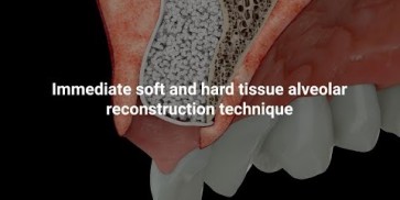
Immediate tissue augmentation technique with application of cerabone® and Jason® membrane
Ridge preservation by Dr. Derk Siebers
Lateral augmentation on pig jaw by PD Dr. Dr. D. Rothamel
GBR with cerabone® and Jason® membrane – Dr. Alfonso Caiazzo (Italy)
cerabone® - natural bovine bone grafting material
cerabone® - excellent biofunctionality - superior hydrophilicity and blood uptake

La pronunciada hidrofilia de la superficie de cerabone® produce una absorción rápida de sangre o suero salino, lo que mejora sus propiedades de manejo. Igualmente, su entramado poroso tridimensional permite una penetración y absorción rápida de las proteínas sanguíneas y séricas y actúa como reservorio para proteínas y factores de crecimiento. Su exclusivo proceso de manufacturación basado en calentamiento a altas temperaturas elimina todos los componentes orgánicos y potencialmente antigénicos, convirtiéndolo en un material seguro y libre de proteínas. cerabone® es un material de injerto óseo natural bovino que es el material preferido para un elevado número de dentistas. Hasta 2016, más de 650.000 pacientes en más de 90 países han sido tratados con éxito con cerabone®.

[1] Tadic, D. and Epple, M. Biomaterials 2004; Vol. 25 No. 6, pp. 987–994