cerabone® and mucoderm® for immediacy in esthetic zone -Dr. M Motta
-
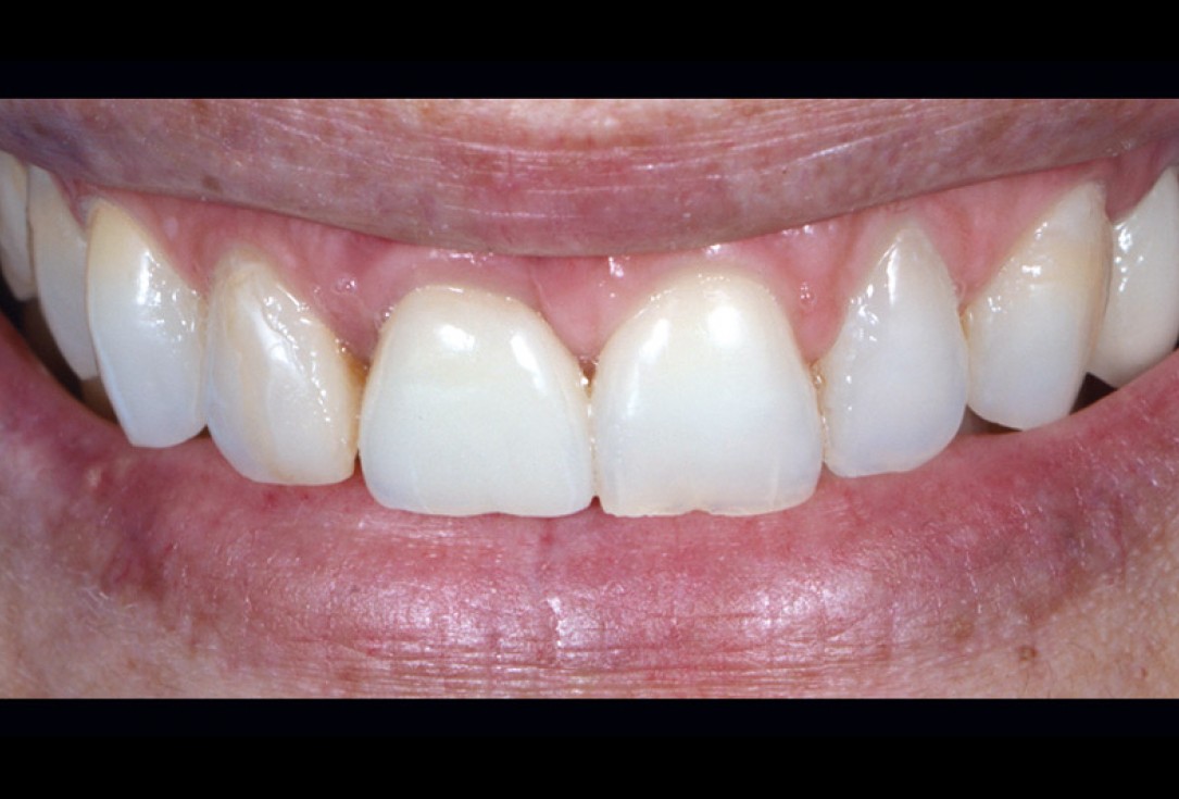
1/18 - Initial view of the case. Discoloration of 1.1 and mild class I gingival recessioncerabone® and mucoderm® for immediacy in esthetic zone -Dr. M Motta
-
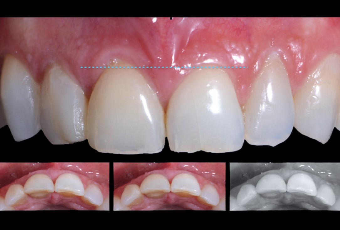
2/18 - Frontal view of the case. Suspicion of root fracture. Changes in occlusal line and extrusion of 1.1cerabone® and mucoderm® for immediacy in esthetic zone -Dr. M Motta
-
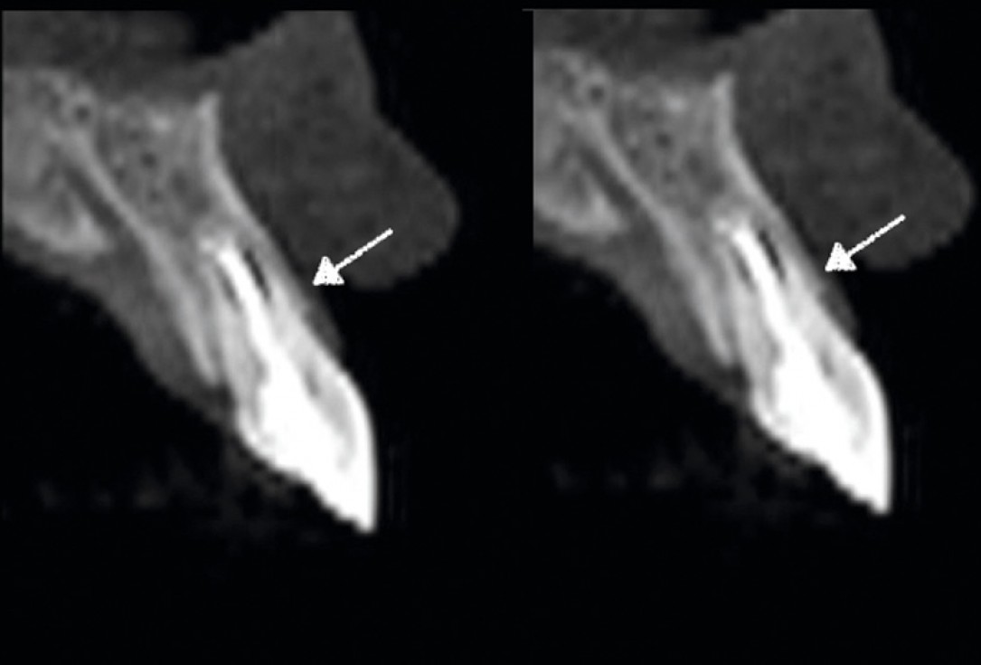
3/18 - CT confirming the suspected Dx.cerabone® and mucoderm® for immediacy in esthetic zone -Dr. M Motta
-
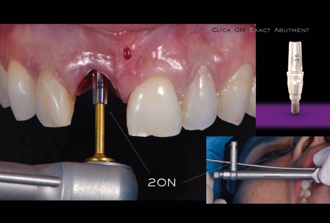
4/18 - Immediate placement of Neodent Helix GM Acqua implant.cerabone® and mucoderm® for immediacy in esthetic zone -Dr. M Motta
-
5/18 - Intrasurgical view shows implant with good stability and a bone gap between implant and vestibular platecerabone® and mucoderm® for immediacy in esthetic zone -Dr. M Motta
-
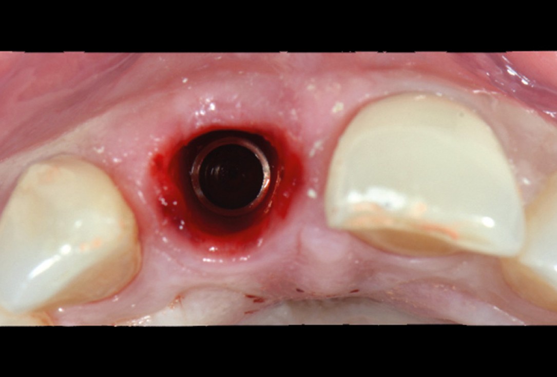
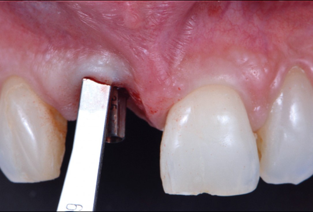
6/18 - Placement of definitive abutmentcerabone® and mucoderm® for immediacy in esthetic zone -Dr. M Motta
-
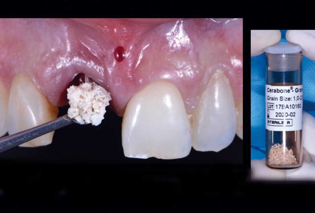
7/18 - Filling bone gap with cerabone® granules.cerabone® and mucoderm® for immediacy in esthetic zone -Dr. M Motta
-
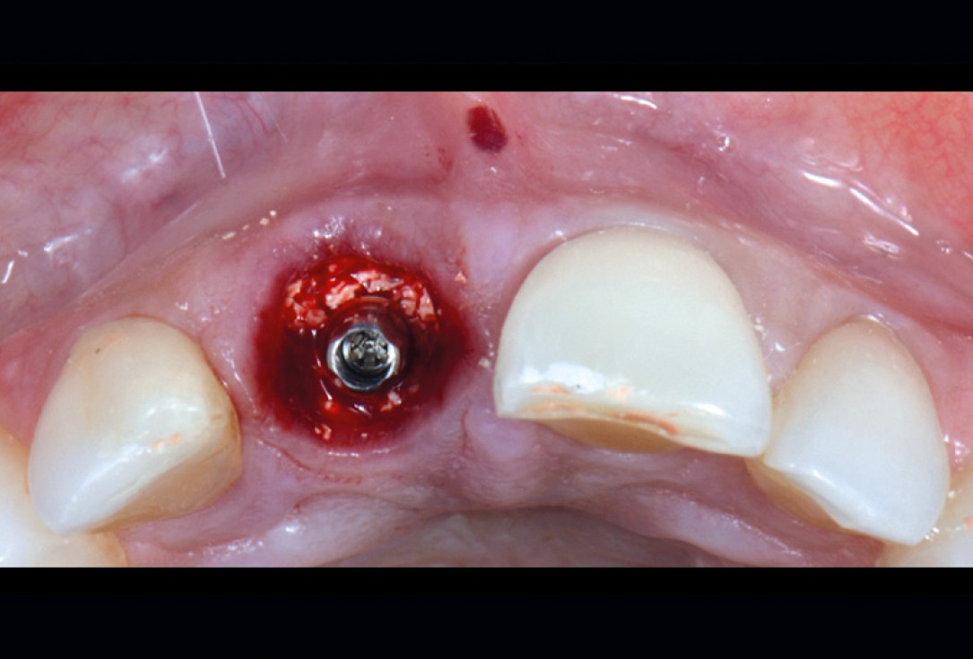
8/18 - Occlusal view of the filled defectcerabone® and mucoderm® for immediacy in esthetic zone -Dr. M Motta
-
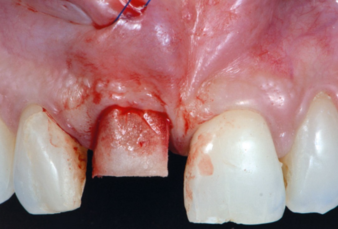
9/18 - Preparation of a gingival envelop flapcerabone® and mucoderm® for immediacy in esthetic zone -Dr. M Motta
-
10/18 - Fixation of mucoderm® to the bottom of the envelop using a matrix suture.cerabone® and mucoderm® for immediacy in esthetic zone -Dr. M Motta
-
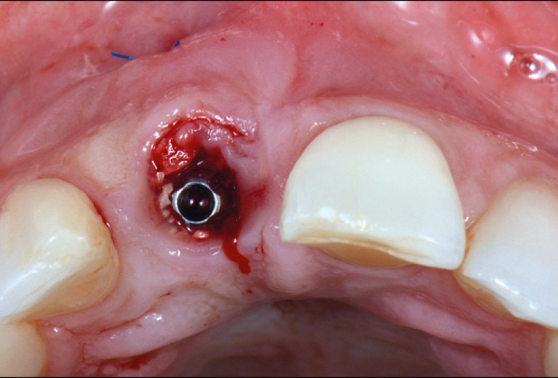
11/18 - Oclussal view of the placed mucoderm® and cerabone®.cerabone® and mucoderm® for immediacy in esthetic zone -Dr. M Motta
-
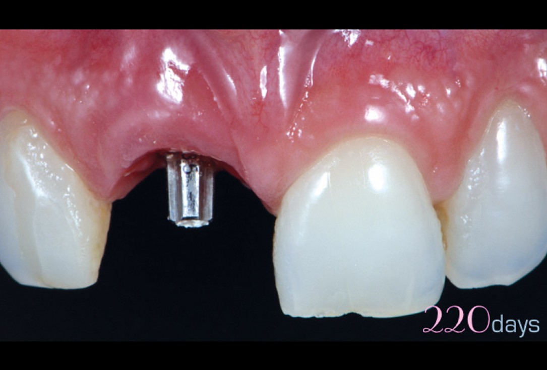
12/18 - Clinical view 220 days post-surgery.cerabone® and mucoderm® for immediacy in esthetic zone -Dr. M Motta
-
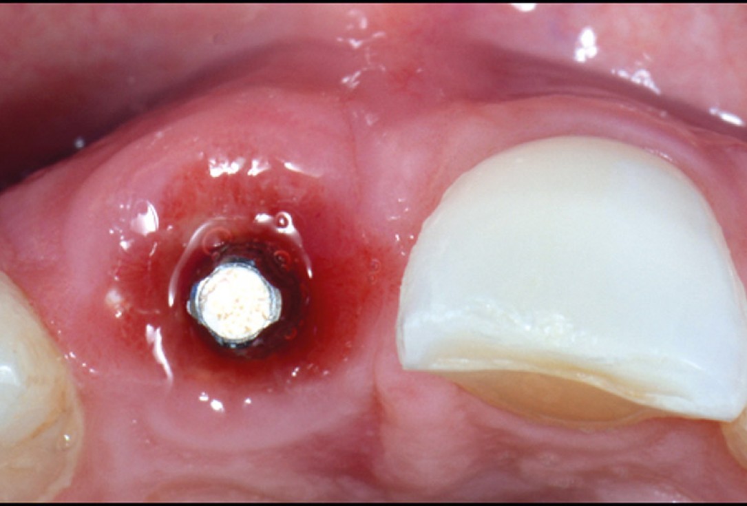
13/18 - Oclussal post-surgical viewcerabone® and mucoderm® for immediacy in esthetic zone -Dr. M Motta
-
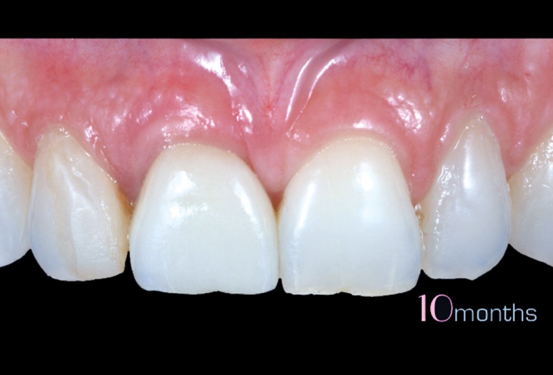
14/18 - Clinical view 10 months after the final restorationcerabone® and mucoderm® for immediacy in esthetic zone -Dr. M Motta
-
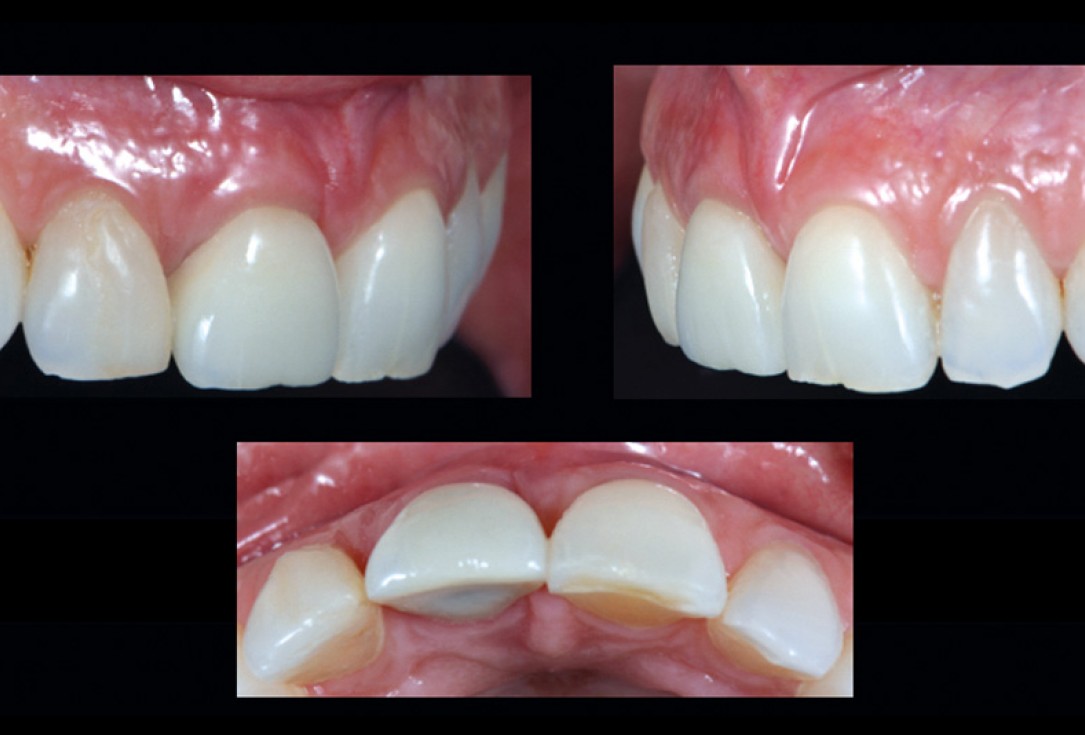
15/18 - Lateral and occlusal view of the final restaurationcerabone® and mucoderm® for immediacy in esthetic zone -Dr. M Motta
-
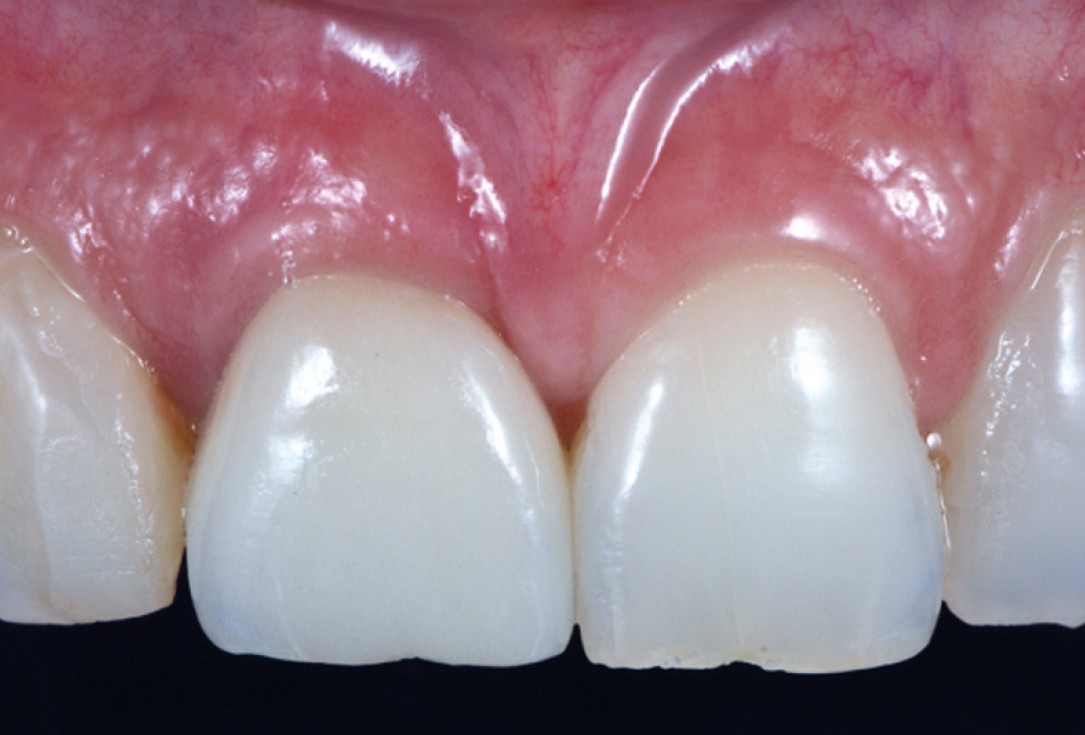
16/18 - Close-up of the final case viewcerabone® and mucoderm® for immediacy in esthetic zone -Dr. M Motta
-
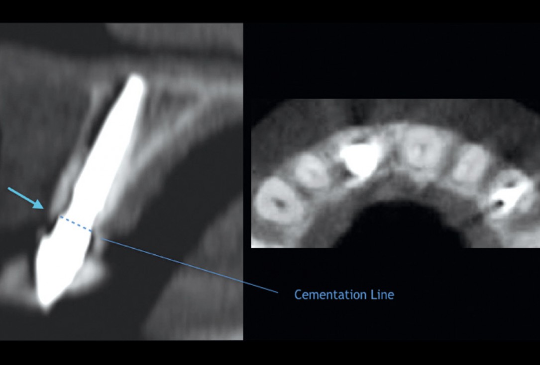
17/18 - Control CT and occlusal Rx.cerabone® and mucoderm® for immediacy in esthetic zone -Dr. M Motta
-
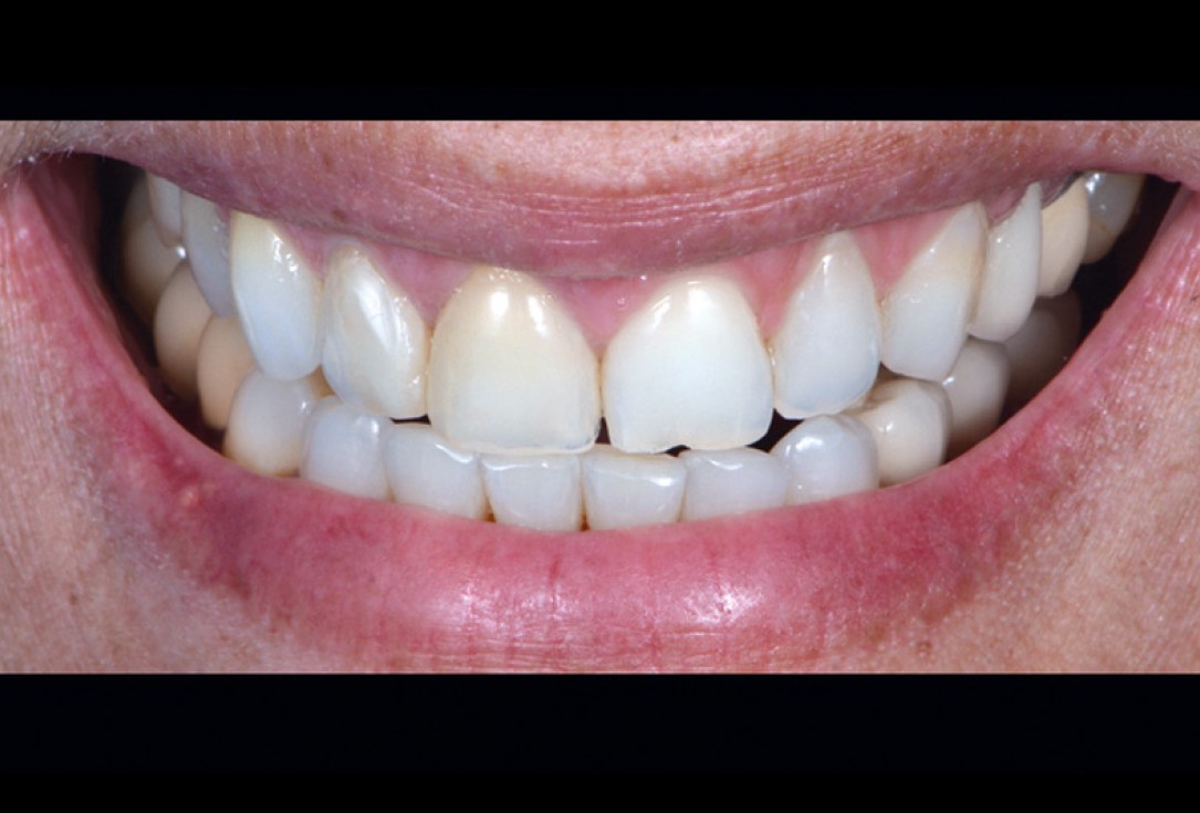
18/18 - Natural smile of the patientcerabone® and mucoderm® for immediacy in esthetic zone -Dr. M Motta

Initial clinical situation. Atrophic maxillary ridge.

Initial x-ray showing bone loss around implants placed 5 years ago in another dental clinic

Initial situation: missing teeth #11 & 12 and badly broken #21 root

Pre-operative OPG shows deep vertical intrabony defects on the distal aspects of teeth 13 and 14.

Instable bridge situation with abscess formation at tooth #15 after apicoectomy

Initial clinical situation.

Implant insertion in atrophic alveolar ridge

Preoperative clinical situation

Pre-operative OPG

Initial clinical situation.

Pre-surgical situation.

Pre-operative X-ray. Hopless tooth 21.

Pre-surgical situation. Teeth 26 and 27 missing.

Extraction of tooth 21 after endodontic treatment

Pre-surgical probing reveals a deep intrabony defect on the distal aspect of the upper canine.

Initial clinical situation with single tooth gap in regio 21

Pre-operative radiographic view. Intrabony defect on the distal aspect of the lateral incisor.

Clinical situation before extraction and implantation

Pre-operative radiographic view.

Situation after tooth extraction.

Situation after tooth removal.

Initial clinical situation with gum recession and labial bone loss eight weeks following tooth extraction

Three implants placed in a narrow posterior mandible

Pre-operative clinical situation.

Clinical situation with narrow alveolar ridge in the lower jaw

Initial clinical situation showing bone wall defect.
