Horizontal GBR using permamem®, cerabone® and maxgraft® granules and soft tissue augmentation with mucoderm® - Dres. H. Maghaireh and V. Ivancheva
-
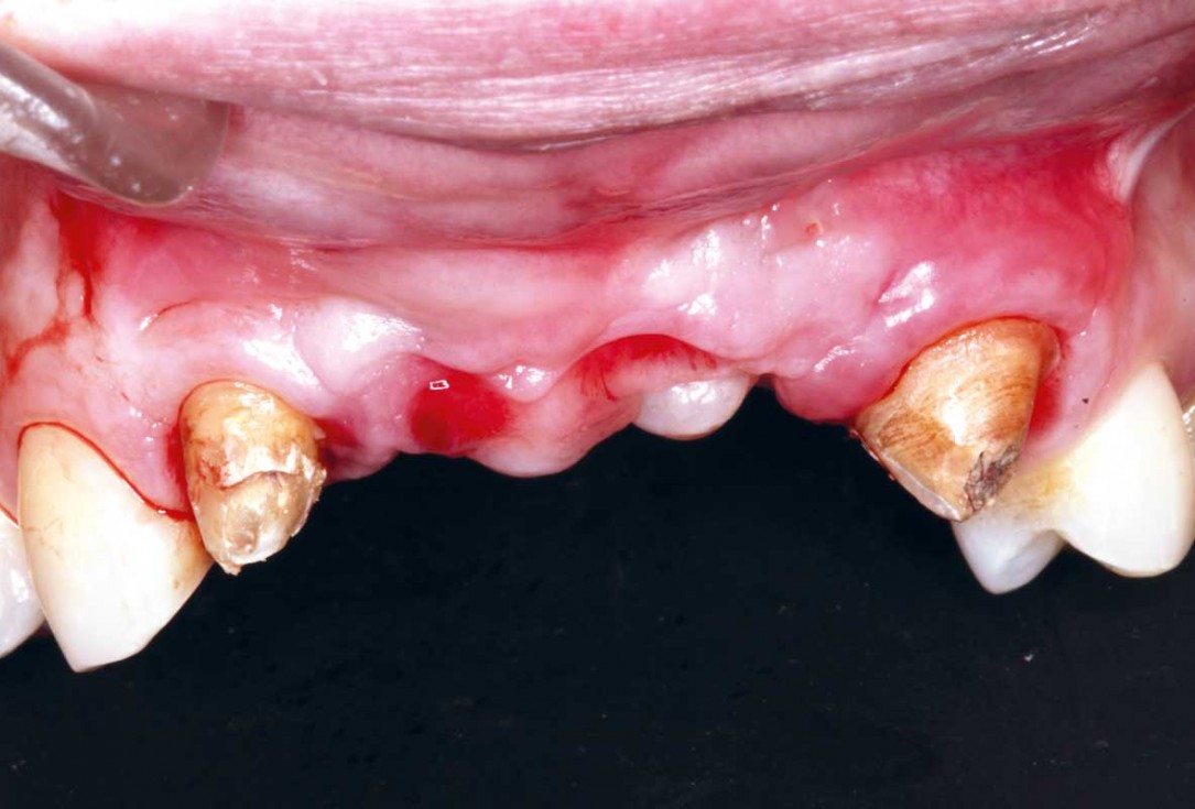
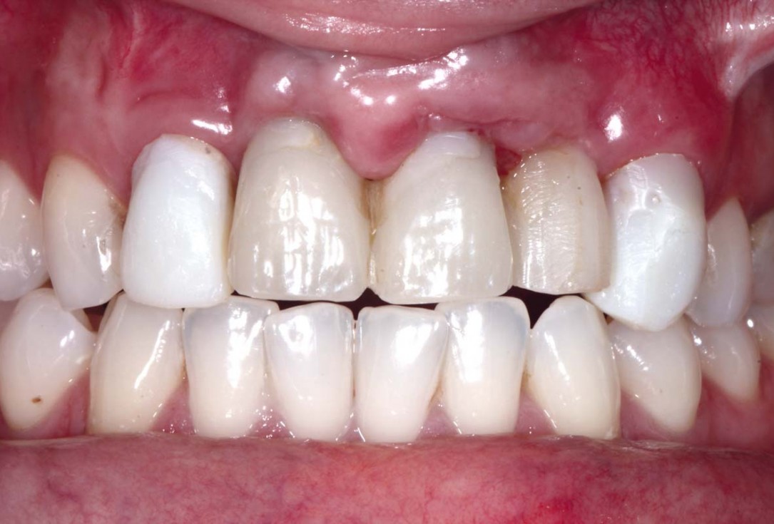
01/33 - Initial situation - A young female 34 years old lost her front teeth in an surfing accident and she had a 5 unit bridge supported by her upper left lateral and right canine. The restoration failed and both supporting crowns have exposed and leaking margins.Horizontal GBR using permamem®, cerabone® and maxgraft® granules and soft tissue augmentation with mucoderm® - Dres. H. Maghaireh and V. Ivancheva
-
02/33 - Initial situation - A young female 34 years old lost her front teeth in an surfing accident and she had a 5 unit bridge supported by her upper left lateral and right canine. The restoration failed and both supporting crowns have exposed and leaking margins.Horizontal GBR using permamem®, cerabone® and maxgraft® granules and soft tissue augmentation with mucoderm® - Dres. H. Maghaireh and V. Ivancheva
-
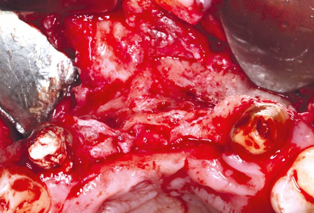
03/33 - Elevating the surgical flap and cleaning the surgical area from the granulation tissueHorizontal GBR using permamem®, cerabone® and maxgraft® granules and soft tissue augmentation with mucoderm® - Dres. H. Maghaireh and V. Ivancheva
-
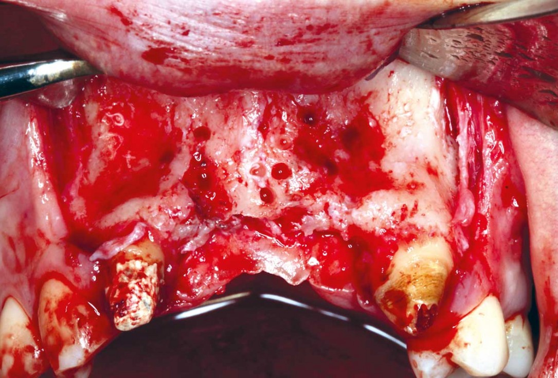
04/33 - Cortectomy was performed with high speed round burrHorizontal GBR using permamem®, cerabone® and maxgraft® granules and soft tissue augmentation with mucoderm® - Dres. H. Maghaireh and V. Ivancheva
-
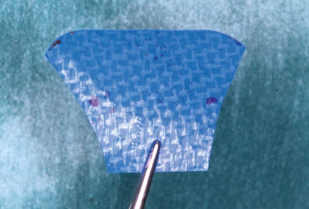
05/33 - Shaping and preparing of permamem®.Horizontal GBR using permamem®, cerabone® and maxgraft® granules and soft tissue augmentation with mucoderm® - Dres. H. Maghaireh and V. Ivancheva
-
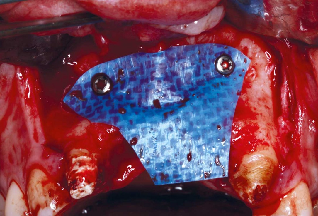
06/33 - Membrane adaptation with fixation screws on the buccal sideHorizontal GBR using permamem®, cerabone® and maxgraft® granules and soft tissue augmentation with mucoderm® - Dres. H. Maghaireh and V. Ivancheva
-
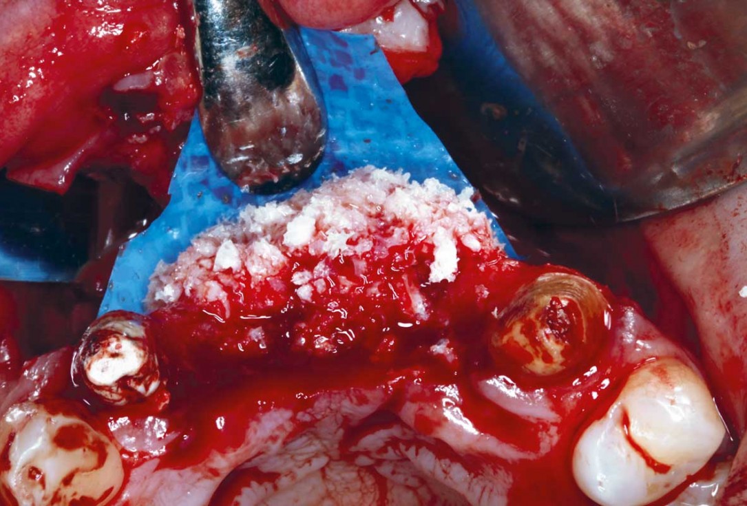
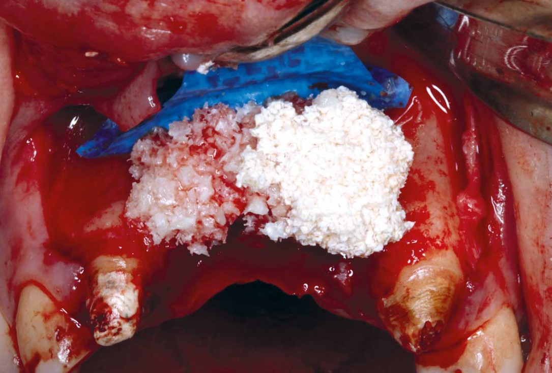
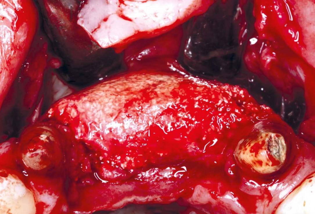
07/33 - Horizontal bone regeneration using layering technique with initial layer of maxgraft® particles mixed with cerabone® (0.5- 1.0 mm)Horizontal GBR using permamem®, cerabone® and maxgraft® granules and soft tissue augmentation with mucoderm® - Dres. H. Maghaireh and V. Ivancheva
-
08/33 - Horizontal bone regeneration using layering technique with initial layer of maxgraft® particles mixed with cerabone® (0.5- 1.0 mm)Horizontal GBR using permamem®, cerabone® and maxgraft® granules and soft tissue augmentation with mucoderm® - Dres. H. Maghaireh and V. Ivancheva
-
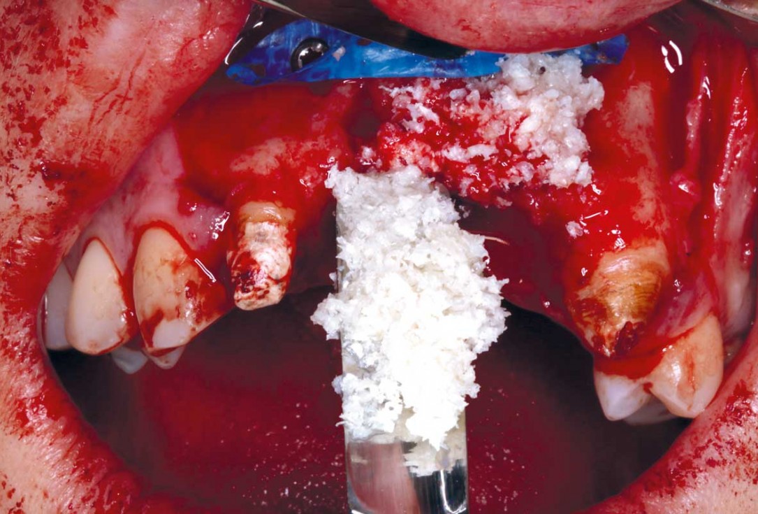
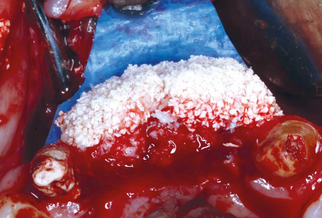
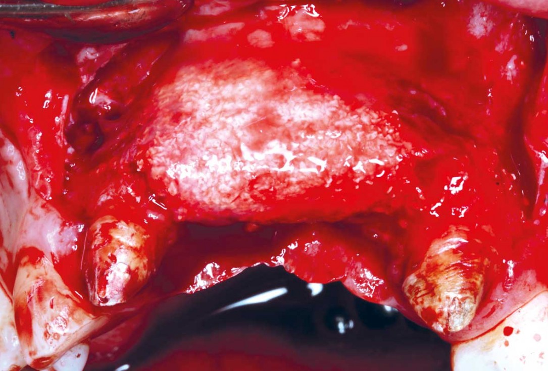
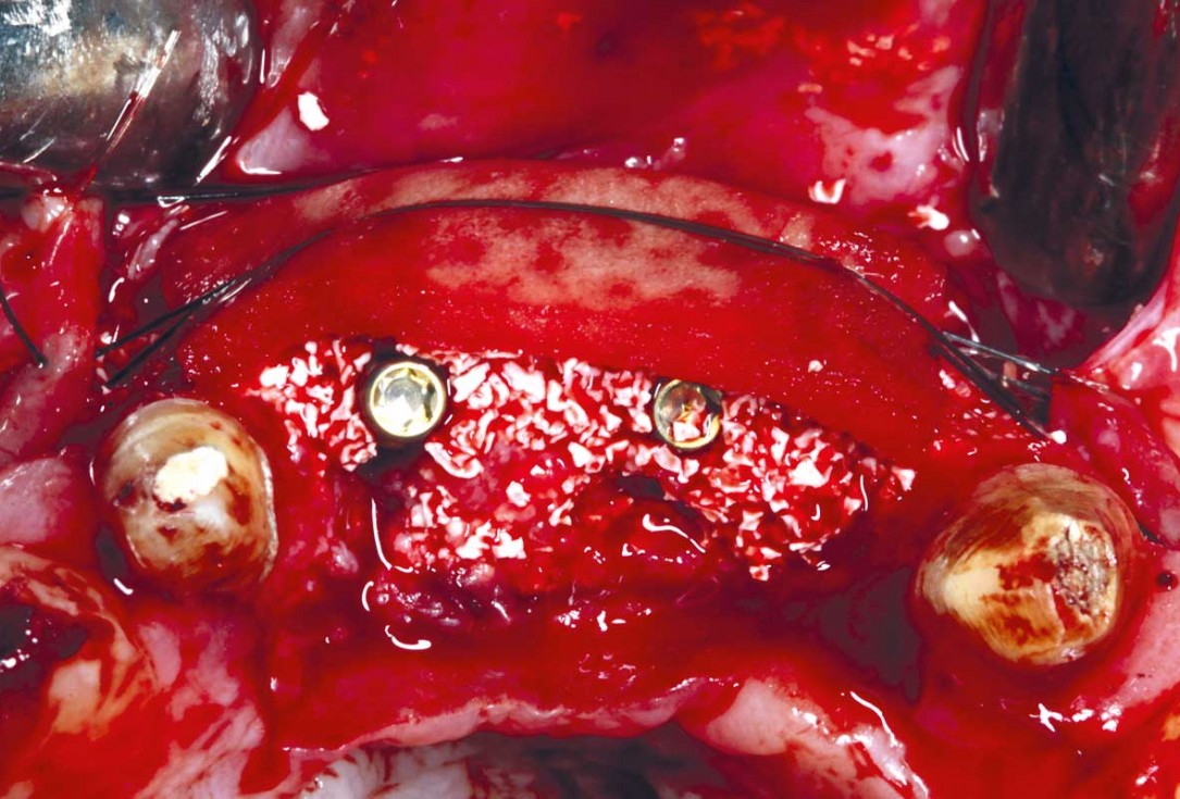
09/33 - Second layer of cerabone® (0.5- 1.0) of 0.5 cc to create convexityHorizontal GBR using permamem®, cerabone® and maxgraft® granules and soft tissue augmentation with mucoderm® - Dres. H. Maghaireh and V. Ivancheva
-
10/33 - Second layer of cerabone® (0.5- 1.0) of 0.5 cc to create convexityHorizontal GBR using permamem®, cerabone® and maxgraft® granules and soft tissue augmentation with mucoderm® - Dres. H. Maghaireh and V. Ivancheva
-
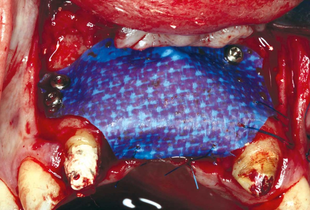
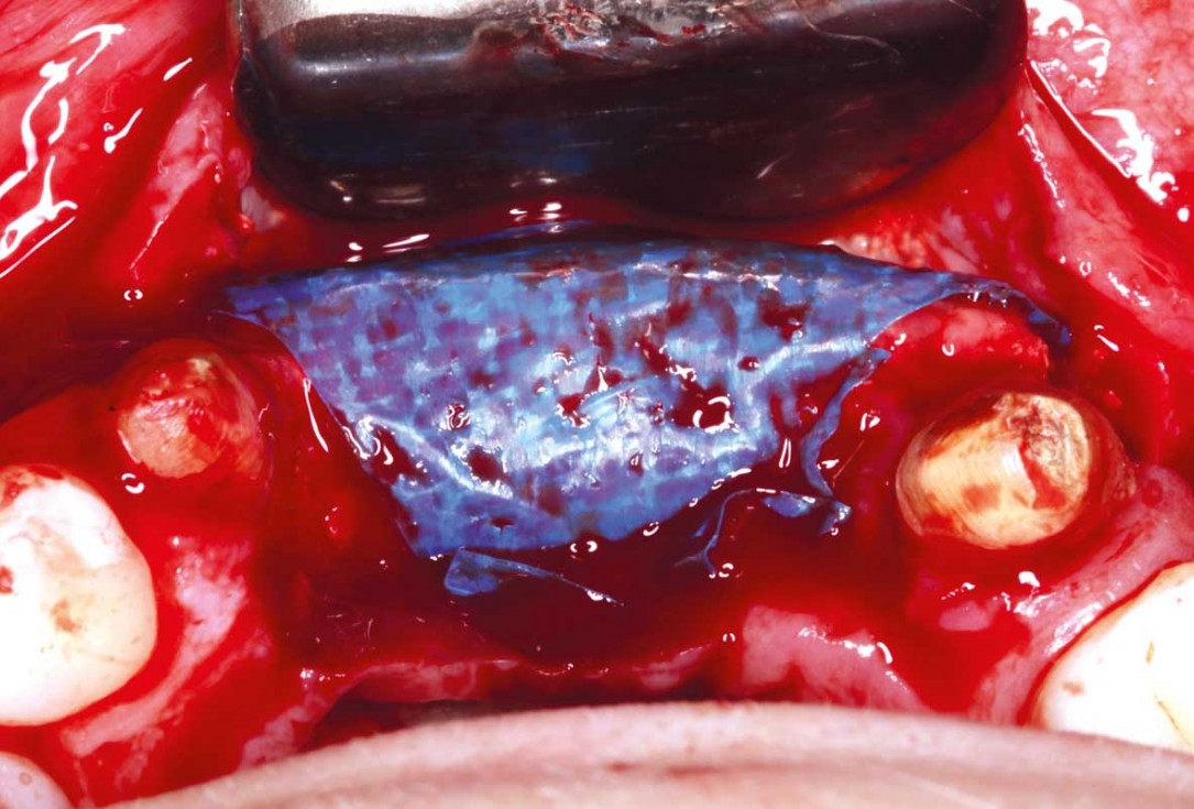
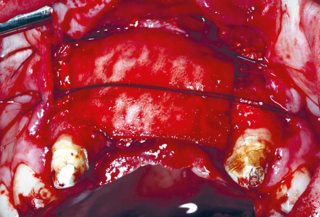
11/33 - Stabilizing the membrane palatal with 4/0 proline sutures.Horizontal GBR using permamem®, cerabone® and maxgraft® granules and soft tissue augmentation with mucoderm® - Dres. H. Maghaireh and V. Ivancheva
-
12/33 - Stabilizing the membrane palatal with 4/0 proline suturesHorizontal GBR using permamem®, cerabone® and maxgraft® granules and soft tissue augmentation with mucoderm® - Dres. H. Maghaireh and V. Ivancheva
-
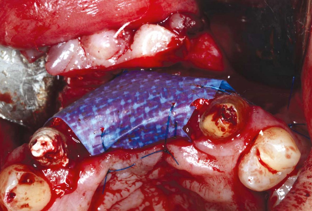
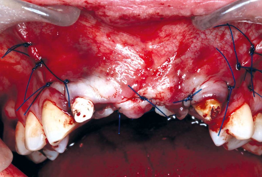
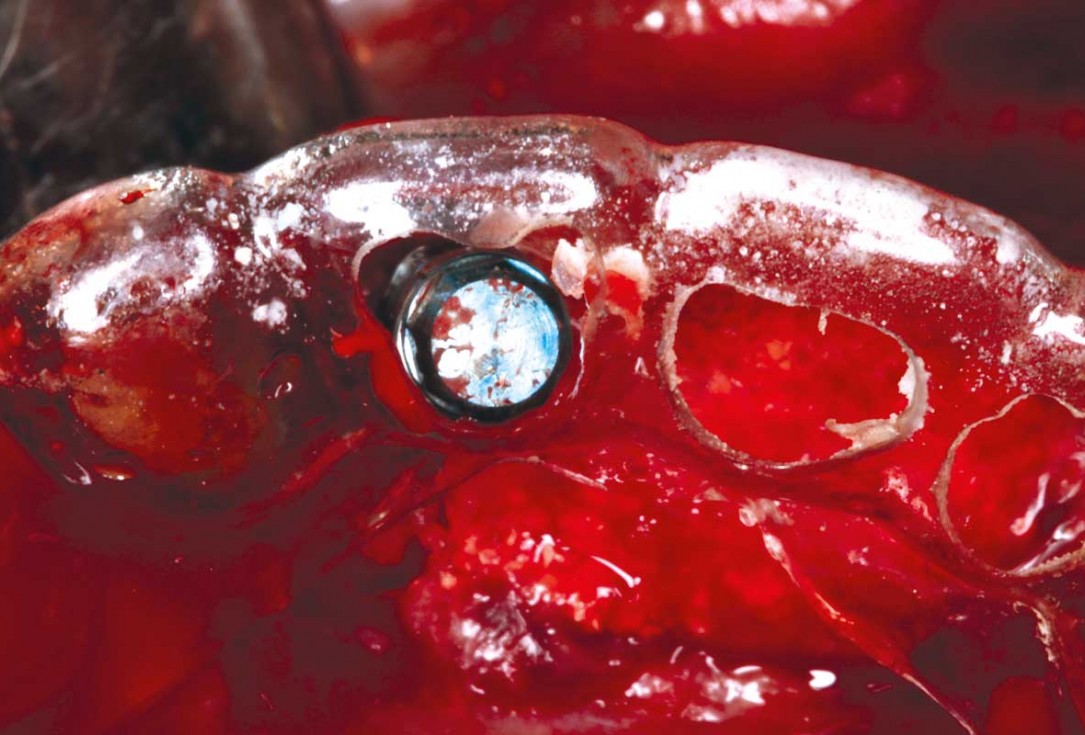
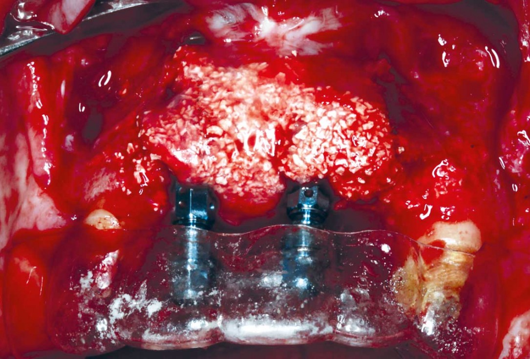
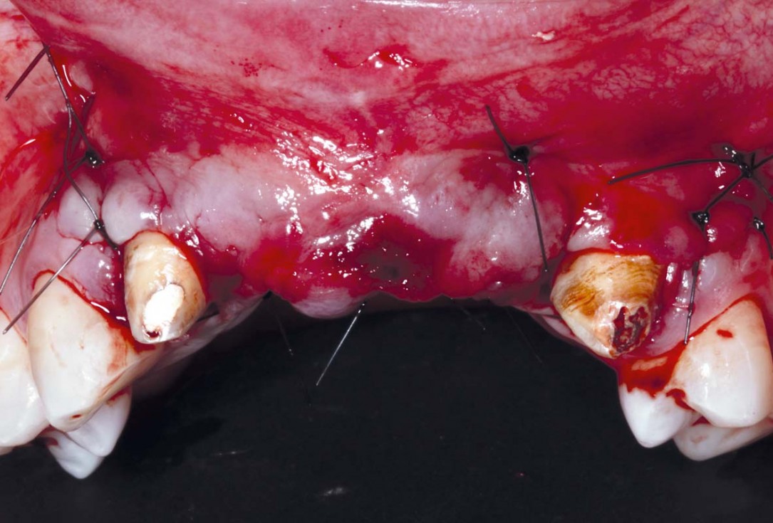
13/33 - Final sutures closed under no tension and fixing and adjusting the temporary bridgeHorizontal GBR using permamem®, cerabone® and maxgraft® granules and soft tissue augmentation with mucoderm® - Dres. H. Maghaireh and V. Ivancheva
-
14/33 - Final sutures closed under no tension and fixing and adjusting the temporary bridgeHorizontal GBR using permamem®, cerabone® and maxgraft® granules and soft tissue augmentation with mucoderm® - Dres. H. Maghaireh and V. Ivancheva
-
15/33 - Initial situation - 24 w after initial horizontal GBR. permamem® is intact and kept the graft in stabile position. Acceptable ridge countering has been achieved and membrane has been removedHorizontal GBR using permamem®, cerabone® and maxgraft® granules and soft tissue augmentation with mucoderm® - Dres. H. Maghaireh and V. Ivancheva
-
16/33 - Initial situation - 24 w after initial horizontal GBR. permamem® is intact and kept the graft in stabile position. Acceptable ridge countering has been achieved and membrane has been removedHorizontal GBR using permamem®, cerabone® and maxgraft® granules and soft tissue augmentation with mucoderm® - Dres. H. Maghaireh and V. Ivancheva
-
17/33 - Stability and density of the bone graft particles after membrane removalHorizontal GBR using permamem®, cerabone® and maxgraft® granules and soft tissue augmentation with mucoderm® - Dres. H. Maghaireh and V. Ivancheva
-
18/33 - Stability and density of the bone graft particles after membrane removalHorizontal GBR using permamem®, cerabone® and maxgraft® granules and soft tissue augmentation with mucoderm® - Dres. H. Maghaireh and V. Ivancheva
-
19/33 - Implant placement in the correct 4D position following the prosthetically driven placement. For central incisions in the cingulum regionHorizontal GBR using permamem®, cerabone® and maxgraft® granules and soft tissue augmentation with mucoderm® - Dres. H. Maghaireh and V. Ivancheva
-
20/33 - Implant placement in the correct 4D position following the prosthetically driven placement. For central incisions in the cingulum regionHorizontal GBR using permamem®, cerabone® and maxgraft® granules and soft tissue augmentation with mucoderm® - Dres. H. Maghaireh and V. Ivancheva
-
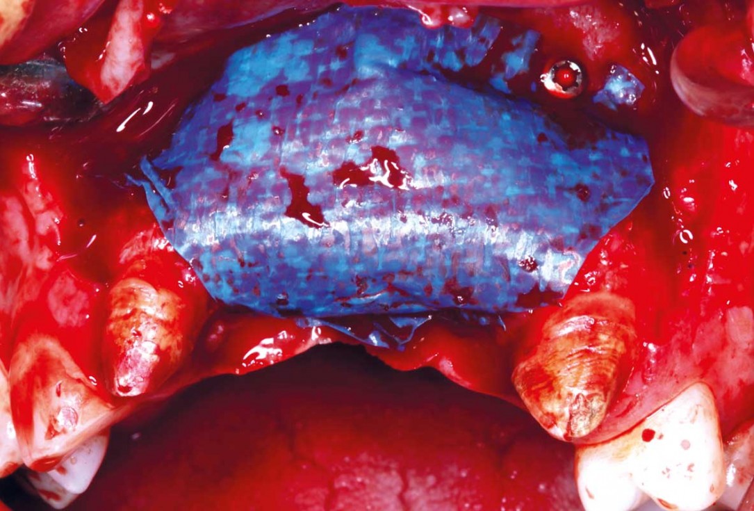
21/33 - Guided bone regeneration after implant placement using cerabone® (0.5- 1.0 mm) shaped to create convexity and covered by two mucoderm® (15x20 mm) used as a soft tissue graft as well as a membrane over the cerabone®. mucoderm® was stabilised with stabilising sutures on each sideHorizontal GBR using permamem®, cerabone® and maxgraft® granules and soft tissue augmentation with mucoderm® - Dres. H. Maghaireh and V. Ivancheva
-
22/33 - Guided bone regeneration after implant placement using cerabone® (0.5- 1.0 mm) shaped to create convexity and covered by two mucoderm® (15x20 mm) used as a soft tissue graft as well as a membrane over the cerabone®. mucoderm® was stabilised with stabilising sutures on each sideHorizontal GBR using permamem®, cerabone® and maxgraft® granules and soft tissue augmentation with mucoderm® - Dres. H. Maghaireh and V. Ivancheva
-
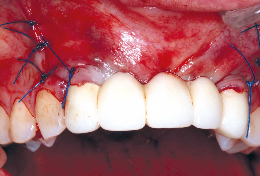
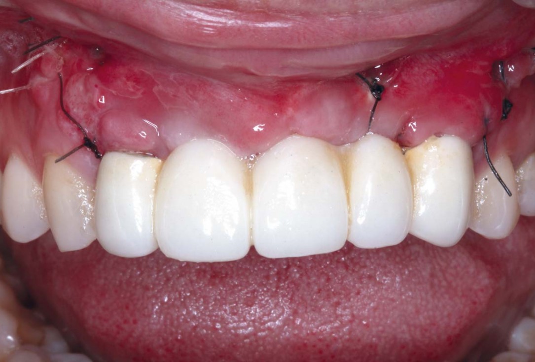
23/33 - Closure sutures and fit of the temporary bridge - cemented on the adjacent peeped teethHorizontal GBR using permamem®, cerabone® and maxgraft® granules and soft tissue augmentation with mucoderm® - Dres. H. Maghaireh and V. Ivancheva
-
24/33 - Closure sutures and fit of the temporary bridge - cemented on the adjacent peeped teethHorizontal GBR using permamem®, cerabone® and maxgraft® granules and soft tissue augmentation with mucoderm® - Dres. H. Maghaireh and V. Ivancheva
-
25/33 - Preparation and fit of fixed screw retained temporary bridge and temporary crowns of the adjacent teeth for soft tissue contouring 18 weeks after implant placementHorizontal GBR using permamem®, cerabone® and maxgraft® granules and soft tissue augmentation with mucoderm® - Dres. H. Maghaireh and V. Ivancheva
-
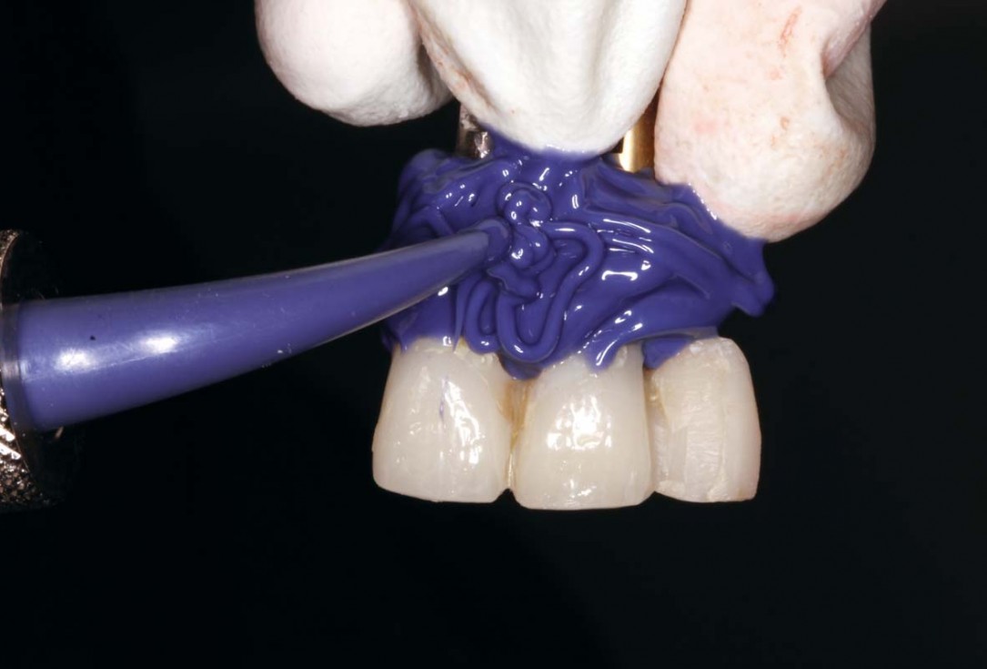
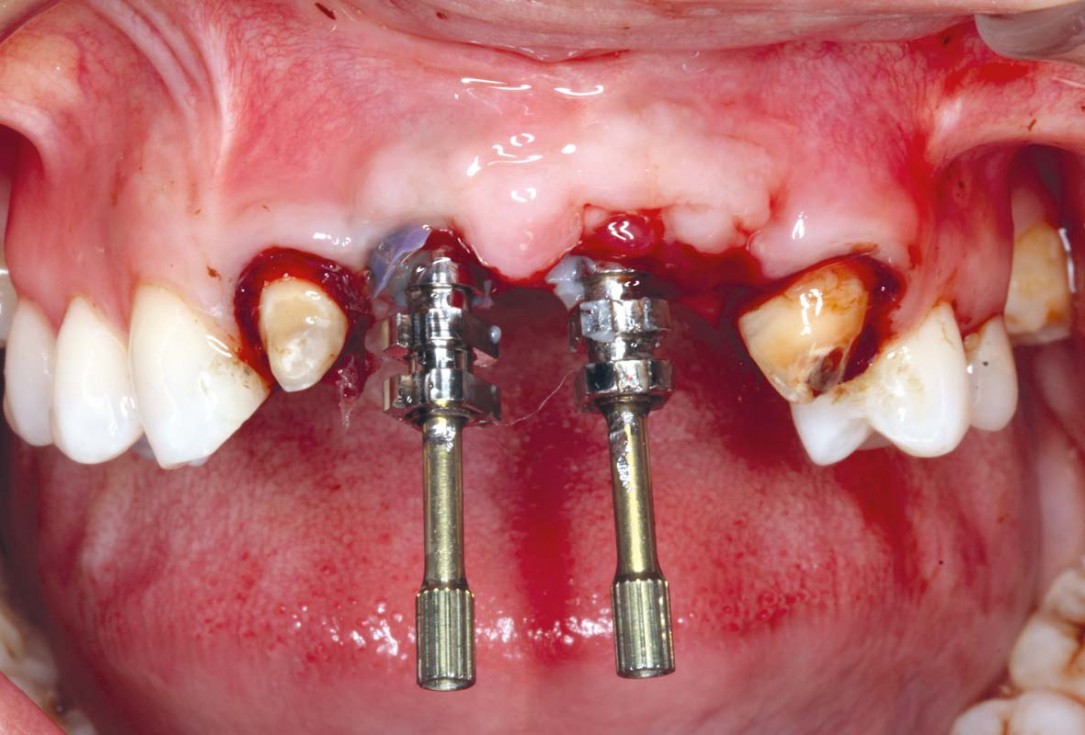
26/33 - Impression and customising the impression pick ups and taking an implant impression for UR1 and UL1 implants with open tray technique 22 weeks after implant placementHorizontal GBR using permamem®, cerabone® and maxgraft® granules and soft tissue augmentation with mucoderm® - Dres. H. Maghaireh and V. Ivancheva
-
27/33 - Impression and customising the impression pick ups and taking an implant impression for UR1 and UL1 implants with open tray technique 22 weeks after implant placementHorizontal GBR using permamem®, cerabone® and maxgraft® granules and soft tissue augmentation with mucoderm® - Dres. H. Maghaireh and V. Ivancheva
-
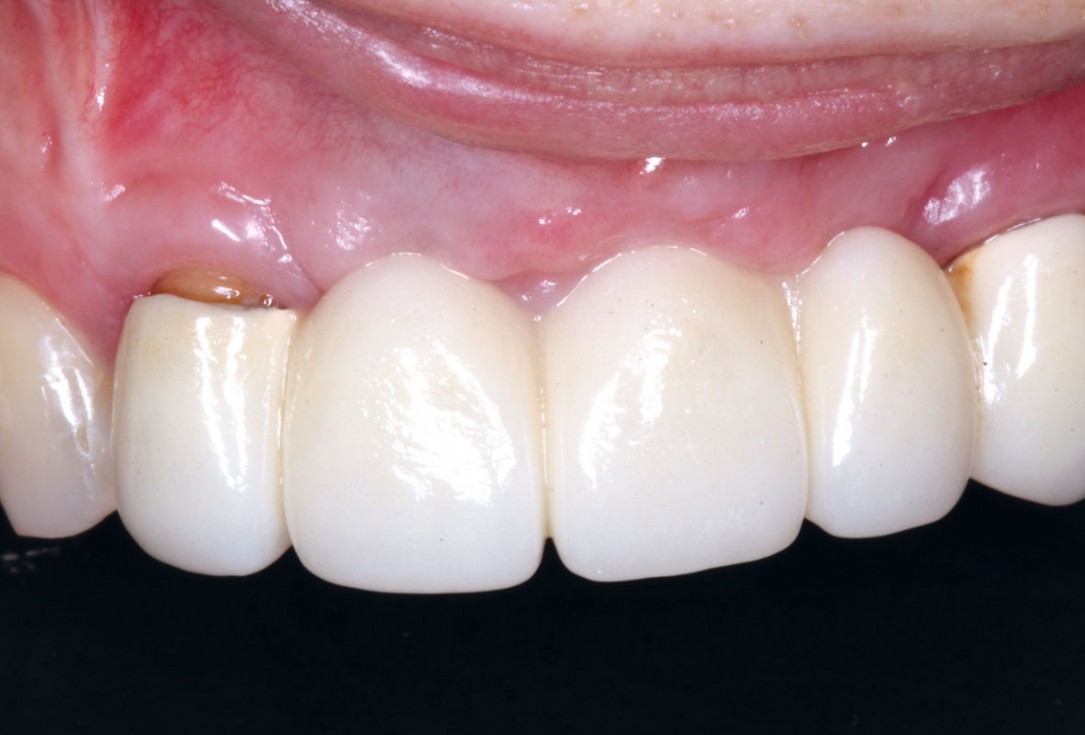
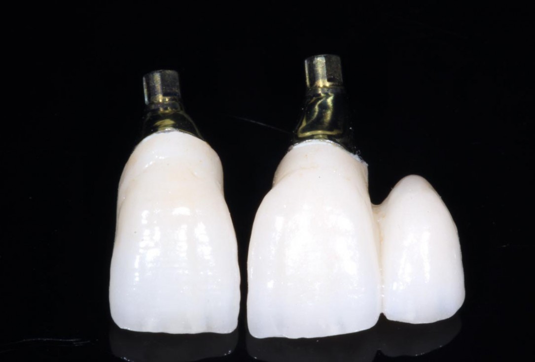
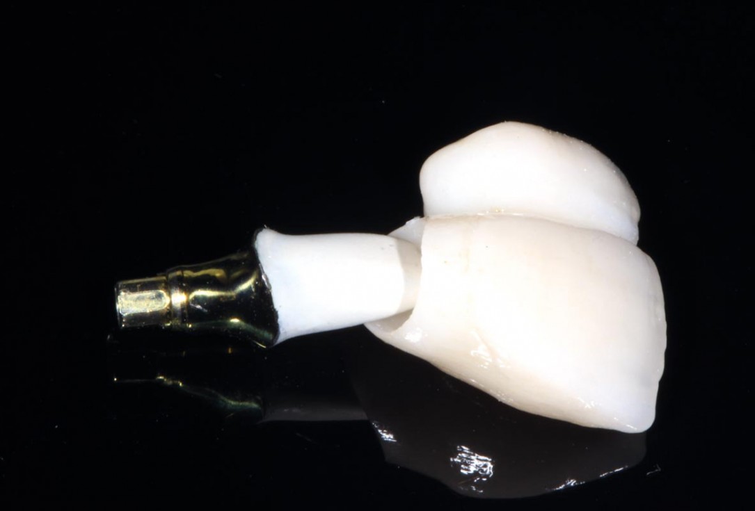
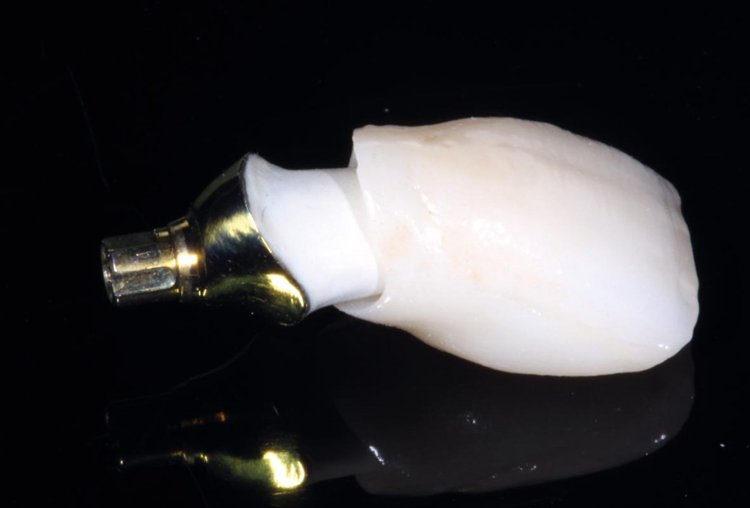
28/33 - Final cement retained porcelain implant crowns on CAD/CAM Ti abutmentsHorizontal GBR using permamem®, cerabone® and maxgraft® granules and soft tissue augmentation with mucoderm® - Dres. H. Maghaireh and V. Ivancheva
-
29/33 - Final cement retained porcelain implant crowns on CAD/CAM Ti abutmentsHorizontal GBR using permamem®, cerabone® and maxgraft® granules and soft tissue augmentation with mucoderm® - Dres. H. Maghaireh and V. Ivancheva
-
30/33 - Final cement retained porcelain implant crowns on CAD/CAM Ti abutmentsHorizontal GBR using permamem®, cerabone® and maxgraft® granules and soft tissue augmentation with mucoderm® - Dres. H. Maghaireh and V. Ivancheva
-
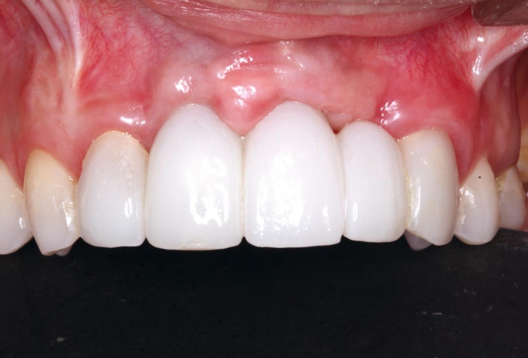
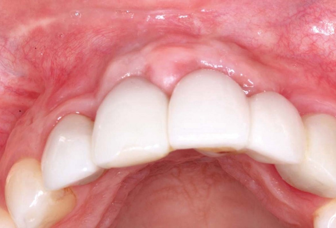
31/33 - Final result 3 months after implant placement surgery and 2 weeks after final prosthetic integration , showing sufficient ridge contouring around the UR1 and UL1 dental implants and satisfactory peri-implant soft tissue thicknessHorizontal GBR using permamem®, cerabone® and maxgraft® granules and soft tissue augmentation with mucoderm® - Dres. H. Maghaireh and V. Ivancheva
-
32/33 - Final result 3 months after implant placement surgery and 2 weeks after final prosthetic integration , showing sufficient ridge contouring around the UR1 and UL1 dental implants and satisfactory peri-implant soft tissue thicknessHorizontal GBR using permamem®, cerabone® and maxgraft® granules and soft tissue augmentation with mucoderm® - Dres. H. Maghaireh and V. Ivancheva
-
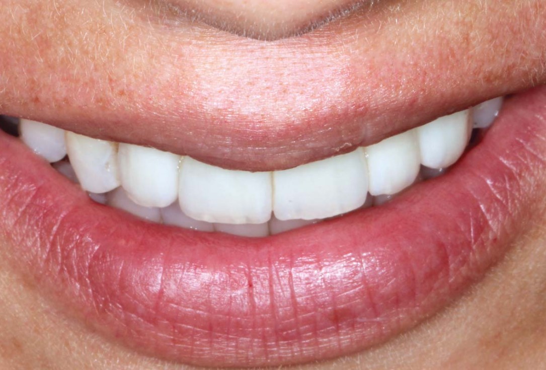
33/33 - Smile line and harmonisation of white and pink aesthetic.Horizontal GBR using permamem®, cerabone® and maxgraft® granules and soft tissue augmentation with mucoderm® - Dres. H. Maghaireh and V. Ivancheva

Initial clinical situation. Atrophic maxillary ridge.

Initial x-ray showing bone loss around implants placed 5 years ago in another dental clinic

Initial view of the case. Discoloration of 1.1 and mild class I gingival recession

Situation after tooth removal.

Initial clinical situation with gum recession and labial bone loss eight weeks following tooth extraction

Three implants placed in a narrow posterior mandible

Pre-operative clinical situation.

Clinical situation with narrow alveolar ridge in the lower jaw

Initial clinical situation showing bone wall defect.

Initial clinical situation.

Pre-surgical situation.

Initial situation: missing teeth #11 & 12 and badly broken #21 root

Pre-operative OPG shows deep vertical intrabony defects on the distal aspects of teeth 13 and 14.

Instable bridge situation with abscess formation at tooth #15 after apicoectomy

Initial clinical situation.

Implant insertion in atrophic alveolar ridge

Preoperative clinical situation

Pre-operative OPG

Pre-operative X-ray. Hopless tooth 21.

Pre-surgical situation. Teeth 26 and 27 missing.

Extraction of tooth 21 after endodontic treatment

Pre-surgical probing reveals a deep intrabony defect on the distal aspect of the upper canine.

Initial clinical situation with single tooth gap in regio 21

Pre-operative radiographic view. Intrabony defect on the distal aspect of the lateral incisor.

Clinical situation before extraction and implantation

Pre-operative radiographic view.
