
mucoderm®
- Augmentation des tissus mous
- Recouvrement des alvéoles d’extraction
- Récessions gingivales
- Greffe de tissus mous en association avec une ROG/RTG
- Élargissement de la gencive attachée
|

Extraction of tooth 44

Pre-surgical clinical situation. Multiple adjacent gingival recessions at teeth 12,13 and 14.

Initial clinical situation. Multiple adjacent gingival recessions in regio 11-13.

Pre-operative clinical situation. Gingival recession at the first premolar.

Insufficient keratinized mucosa and extremely shallow vestibule on the maxilla

Initial clinical situation

Initial clinical situation

Initial clinical situation with lack of keratinized tissue

Lack of sufficient keratinized mucosa following extensive horizontal ridge augmentation

Pre-operative clinical situation. Multiple adjacent gingival recessions.

Longitudinal fracture on the root resected tooth 21 with visible buccal fistula


Tooth extraction due to root fracture

Drilling template for guided implant placement


Clinical situation before extraction

Initial situation: missing teeth #11 & 12 and badly broken #21 root

Initial clinical situation with Miller class 1 recession

Initial view of the case. Discoloration of 1.1 and mild class I gingival recession

Preoperative situation – Maxillary defect in area 14-16 (loss of implant 16 due to periimplantitis, tooth 14 extracted recently and area 15 already edentulous for a while)

Initial clinical situation shows an odontogenic fibroma that was growing for years

Initial clinical situation with pronounced vertical and horizontal bone defect

Initial clinical situation - Central incisors with dental destruction and periapical pathology

Situation before extraction of the teeth


Multiple adjacent gingival recessions.

Pre-surgical situation. Multiple adjacent gingival recessions at teeth 12, 13 and 14.


Pre-operative clinical view. Multiple adjacent gingival recessions.

Initial clinical situation with narrow ridge

Pre-operative clinical situation. Gingival recessions at teeth 11 and 21.

Initial clinical situation

X-ray shows a 3-dimensional periondontal defect

Clinical situation before surgery

Baseline clinical situation, frontal view.

X-ray showing endodontic failure of the molar

Initial clinical situation showing severe soft tissue loss

Alveolar socket before soft and hard tissue augmentation

Clinical situation

Initial clinical situation showing strongly compromised tooth 21

Initial view of the clinical case: Class III malocclusion
Treatment plan: Regenerative corticotomy (PAOO)

Initial clinical view of the case. Soft tissue dehiscence around implants 26.

Initial situation displaying insufficient bone width


Occlusal view of attached maxgraft® cortico at the buccal site


Initial situation before surgery. Patient lost central incisors 1 month ago due to endodontic failures

Initial clinical situation

Gingival recession at tooth 13. Free gingival graft (FGG) of a previous surgery for root coverage visible.


Initial clinical situation

Pre-surgical clinical situation. Deep gingival recessions at both upper canine.

Initial clinical situation with traumatic loss of tooth 21

Pre-operative clinical situation. Shallow multiple adjacent gingival recessions in the first quadrant.

Initial clinical situation

X-ray of initial clinical situation

Multiple adjacent recessions in the upper jaw.

recession on tooth 11

Full-thickness flap preparation bucally and lingually

Intact socket following atraumatic tooth extraction

X-ray control before tooth extraction

Bone defect in area 11-21 due to two lost implants (periimplantitis) after 15 years of function


Pre-operative clinical situation: changed color in the gingiva in the front maxilla

Initial clinical situation showing tooth 45 not worth preserving

Initial situation - A young female 34 years old lost her front teeth in an surfing accident and she had a 5 unit bridge supported by her upper left lateral and right canine. The restoration failed and both supporting crowns have exposed and leaking margins.


Clinical view 8 weeks after extraction of teeth 25 and 26

- Revascularisation et intégration rapides
- Remplacement des tissus mous sans prélèvement d’un greffon autologue palatin
- Remodelage complet dans les propres tissus du patient
- Temps de résorption d’environ 6 à 9 mois
- Mise en place et fixation aisées
- Possibilité de découpe pour des procédures spécifiques
- Épaisseur d’environ 1,2-1,7 mm
Art. -No. | Size | Content |
|---|---|---|
701520 | 15x20 | 1 matrix |
702030 | 20x30 | 1 matrix |
703040 | 30x40 | 1 matrix |

Après la mise en place, le sang du patient s’infiltre dans la greffe de mucoderm® à travers le réseau tridimensionnel des tissus mous, en amenant des cellules hôtes à la surface de la greffe de tissus mous et en déclenchant le processus de revascularisation. Une revascularisation importante peut débuter après l’implantation, en fonction de l’état de santé du patient. mucoderm® constitue une alternative sûre à la greffe autologue de tissu conjonctif, et convient à une large palette d’indications de greffe de tissus mous.
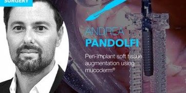
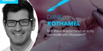
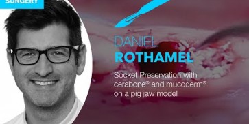
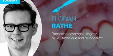
Please find our free webinars at www.botiss-webinars.com
Kostenfreie Webinare zu Schulungszwecken finden Sie unter www.botiss-webinars.com
Please find our free webinars at www.botiss-webinars.com
Please find our free webinars at www.botiss-webinars.com
Please find our free webinars at www.botiss-webinars.com
Please find our free webinars at www.botiss-webinars.com
Please find our free webinars at www.botiss-webinars.com
Please Contact us for Literature.


