Ridge preservation with maxgraft®, mucoderm® & Jason® membrane - Dr. F. Rojas-Vizcaya
-
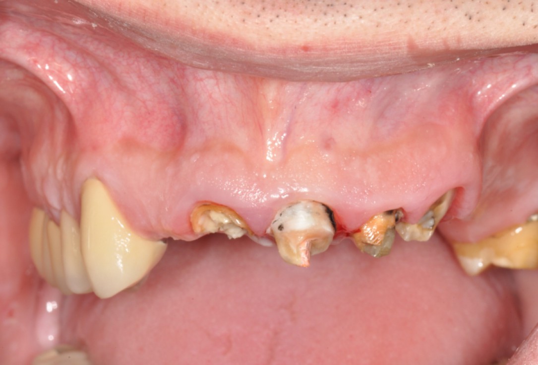
01/19 - Situation before extraction of the teethRidge preservation with maxgraft®, mucoderm® & Jason® membrane - Dr. F. Rojas-Vizcaya
-
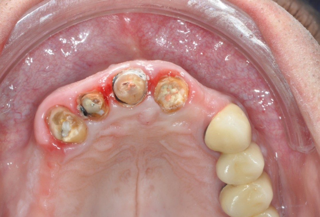
02/19 - Situation before extraction of the teeth, occlusal viewRidge preservation with maxgraft®, mucoderm® & Jason® membrane - Dr. F. Rojas-Vizcaya
-
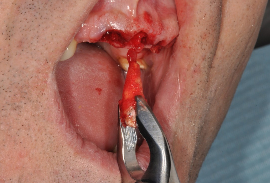
03/19 - Atraumatic tooth extractionRidge preservation with maxgraft®, mucoderm® & Jason® membrane - Dr. F. Rojas-Vizcaya
-
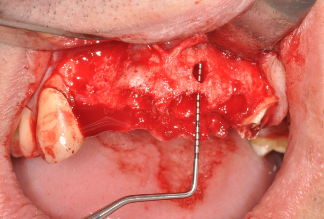
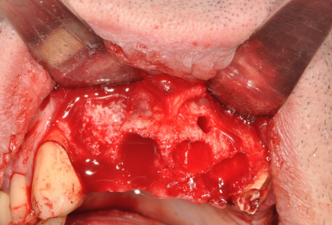
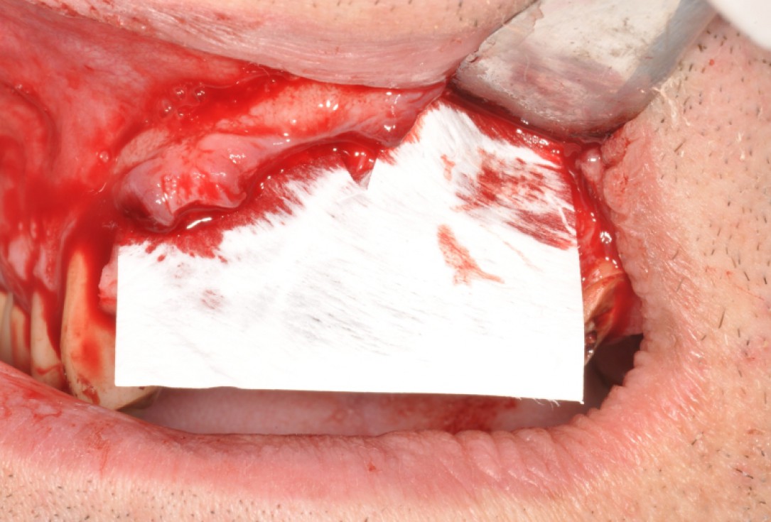
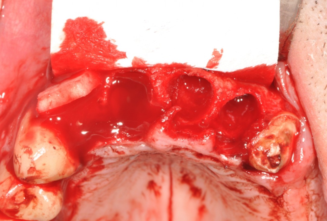
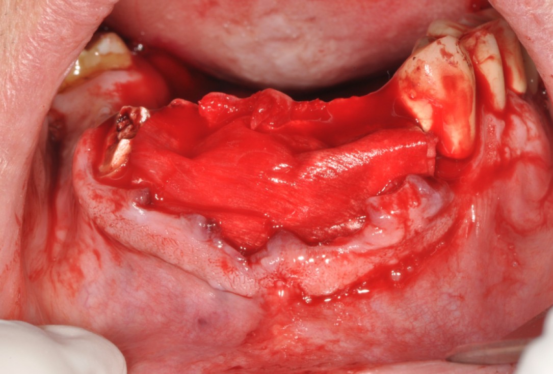
04/19 - Fenestration defect visible after extraction of the teethRidge preservation with maxgraft®, mucoderm® & Jason® membrane - Dr. F. Rojas-Vizcaya
-
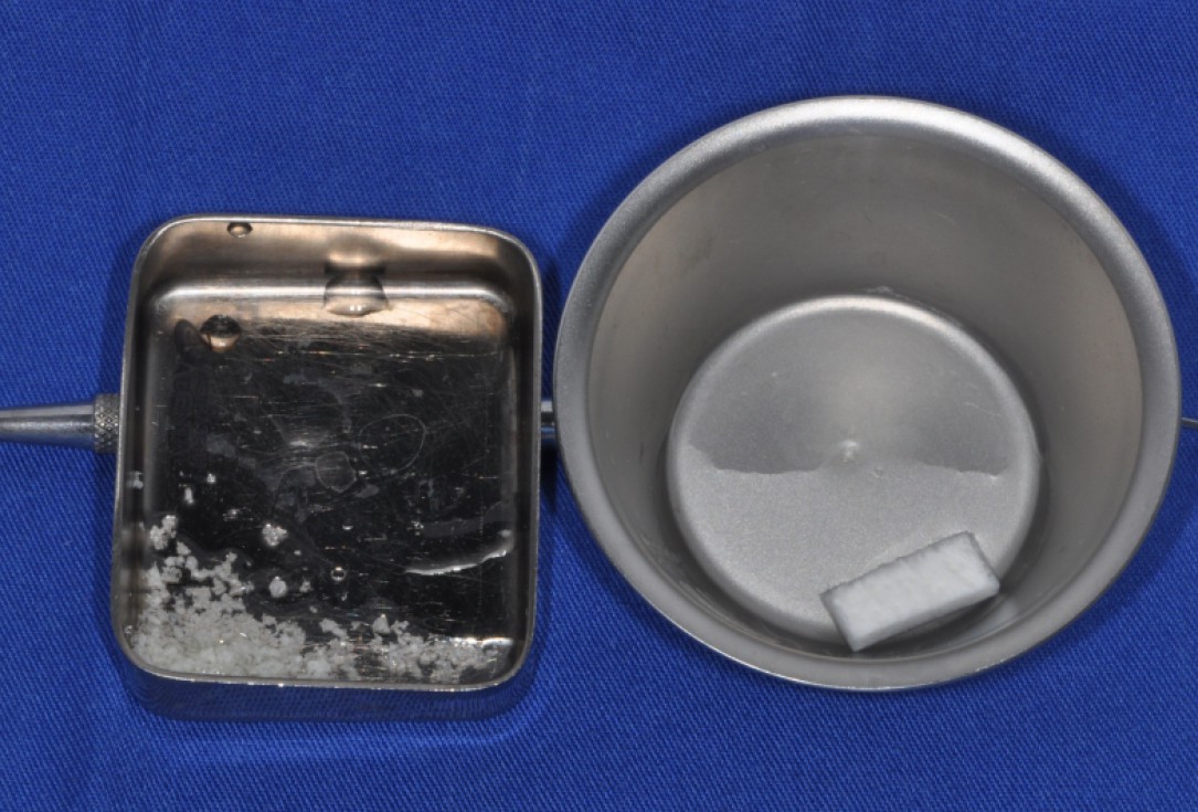
05/19 - Rehydration of mucoderm® and maxgraft® granulesRidge preservation with maxgraft®, mucoderm® & Jason® membrane - Dr. F. Rojas-Vizcaya
-
06/19 - Augmentation od buccal wall in position 12 with maxgraft® granulesRidge preservation with maxgraft®, mucoderm® & Jason® membrane - Dr. F. Rojas-Vizcaya
-
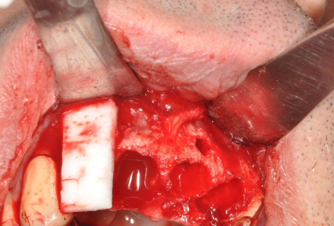
07/19 - Placement of trimmed mucoderm® for soft tissue augmentationRidge preservation with maxgraft®, mucoderm® & Jason® membrane - Dr. F. Rojas-Vizcaya
-
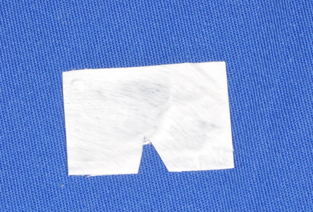
08/19 - Jason® membrane cut to shape for vestibular placementRidge preservation with maxgraft®, mucoderm® & Jason® membrane - Dr. F. Rojas-Vizcaya
-
09/19 - Covering of the vestibular wall with Jason® membraneRidge preservation with maxgraft®, mucoderm® & Jason® membrane - Dr. F. Rojas-Vizcaya
-
10/19 - Jason® membrane protecting the vestibular wallRidge preservation with maxgraft®, mucoderm® & Jason® membrane - Dr. F. Rojas-Vizcaya
-
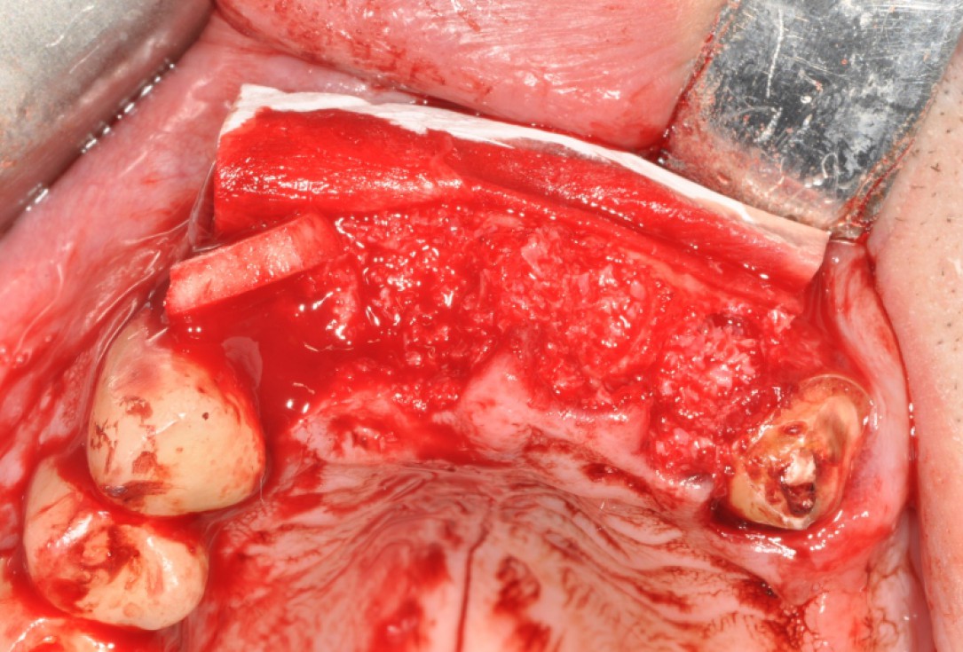
11/19 - Extraction sockets filled with maxgraft® granulesRidge preservation with maxgraft®, mucoderm® & Jason® membrane - Dr. F. Rojas-Vizcaya
-
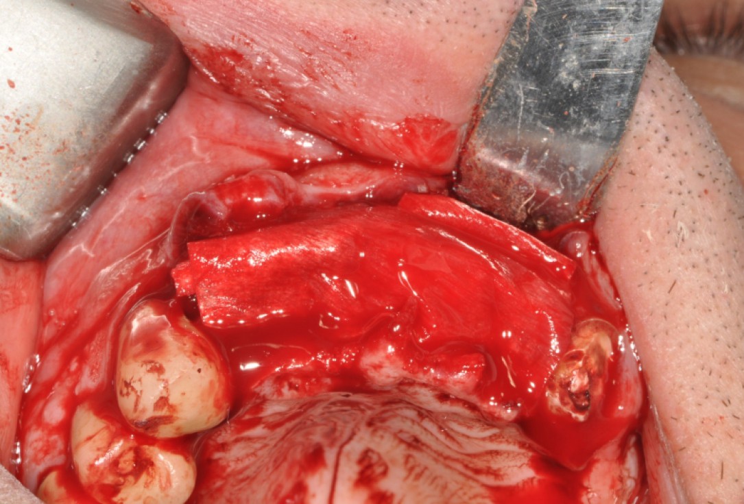
12/19 - Jason® membrane turned down over the augmented areaRidge preservation with maxgraft®, mucoderm® & Jason® membrane - Dr. F. Rojas-Vizcaya
-
13/19 - Jason® membrane covering the augmented areaRidge preservation with maxgraft®, mucoderm® & Jason® membrane - Dr. F. Rojas-Vizcaya
-
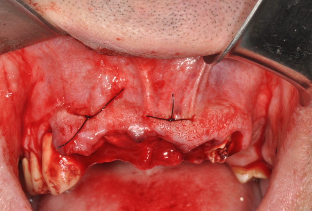
14/19 - Fixation of mucoderm® and Jason® membrane by suturesRidge preservation with maxgraft®, mucoderm® & Jason® membrane - Dr. F. Rojas-Vizcaya
-
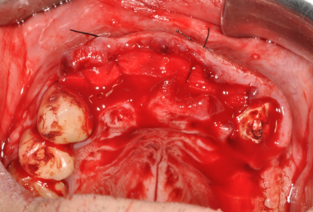
15/19 - Fixation of mucoderm® and Jason® membrane, occlusal viewRidge preservation with maxgraft®, mucoderm® & Jason® membrane - Dr. F. Rojas-Vizcaya
-
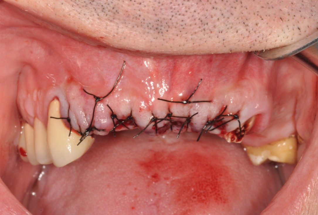
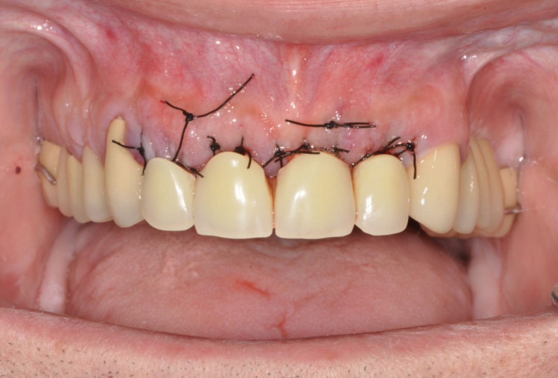
16/19 - Final suturing of the socketsRidge preservation with maxgraft®, mucoderm® & Jason® membrane - Dr. F. Rojas-Vizcaya
-
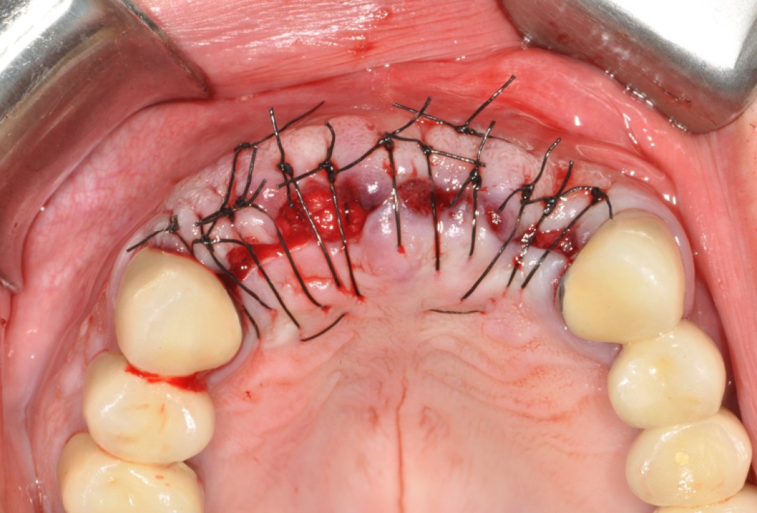
17/19 - Final suturing of the sockets occlusal viewRidge preservation with maxgraft®, mucoderm® & Jason® membrane - Dr. F. Rojas-Vizcaya
-
18/19 - Provisional removable partial denture without compressionRidge preservation with maxgraft®, mucoderm® & Jason® membrane - Dr. F. Rojas-Vizcaya
-
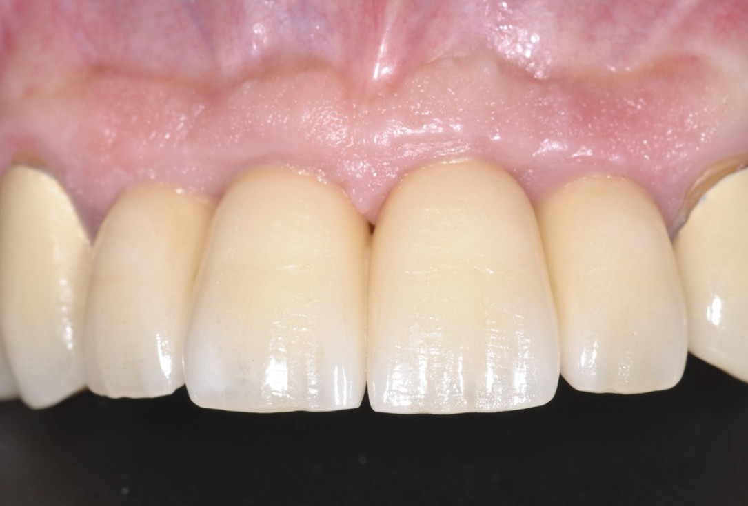
19/19 - Final restoration 7 months post-opRidge preservation with maxgraft®, mucoderm® & Jason® membrane - Dr. F. Rojas-Vizcaya

Situation after tooth removal.

Pre-operative radiographic view.

Three implants placed in a narrow posterior mandible

Initial clinical situation with single tooth gap in regio 21

Initial clinical situation with broken bridge abutment in regio 12 and tooth 21 not worth preserving

Initial clinical situation.

Initial situation 57-year old female patient. X-ray scan reveals severe bone loss due to inflammation in region 13. Treatment plan was extraction of teeth 13 and 14 and augmentation after healing.

X-ray control showing initial situation

Clinical situation

Pre-operative clinical situation: severe atrophy of the maxillary bone

Pre-op picture of affected teeth 11 and 21

Pre-surgical situation. Teeth 26 and 27 missing.

Instable bridge situation with abscess formation at tooth #15 after apicoectomy

Implant insertion in atrophic alveolar ridge

Initial situation after root channel treatment

Initial situation pre-op: Central incisors with mobility 3

Initial clinical situation, regio #16

Lateral view of the defect in the posterior right maxilla.

Preoperative radiological situation

Initial x-ray showing bone loss around implants placed 5 years ago in another dental clinic

Extraction of tooth 21 after endodontic treatment

Initial clinical situation with gum recession and labial bone loss eight weeks following tooth extraction

Preoperative clinical situation

Situation after tooth extraction.

Initial clinical situation

Initial situation after extraction of tooth 21 after 6 months

Bone defect in area 11-21 due to two lost implants (periimplantitis) after 15 years of function

Model of the initial defect computed from a CBCT scan - buccal view
