Soft tissue augmentation and GBR with mucoderm® and maxresorb® - Dr. S. Scherg
-
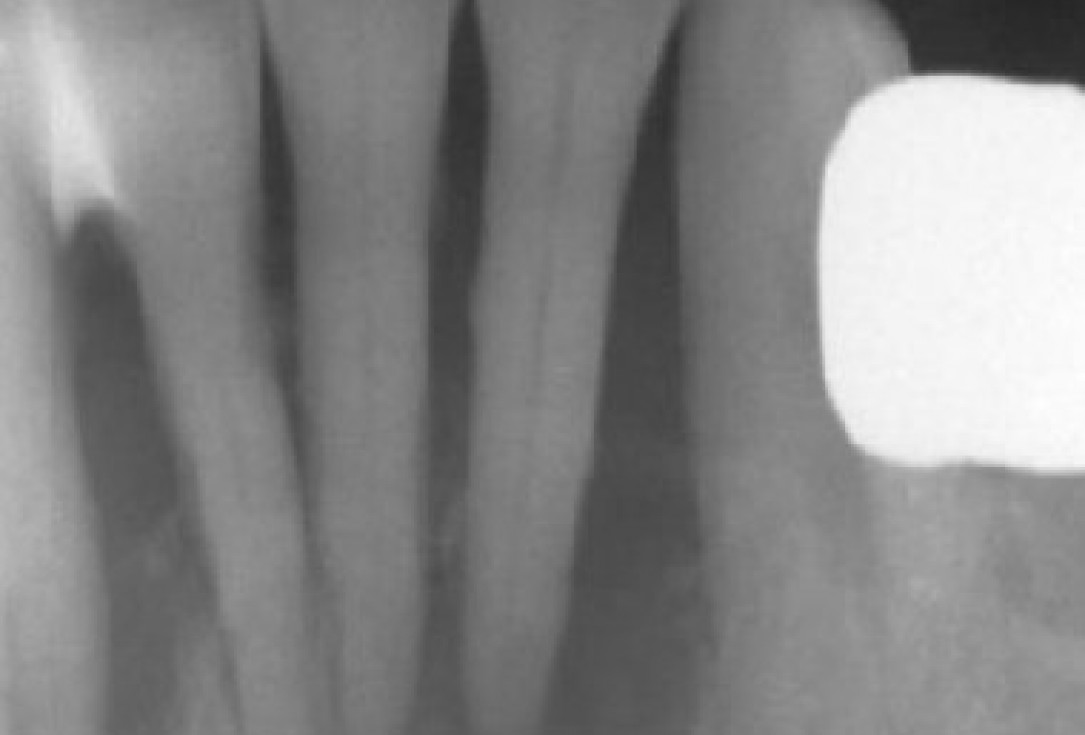
1/15 - X-ray shows a 3-dimensional periondontal defectSoft tissue augmentation and GBR with mucoderm® and maxresorb® - Dr. S. Scherg
-
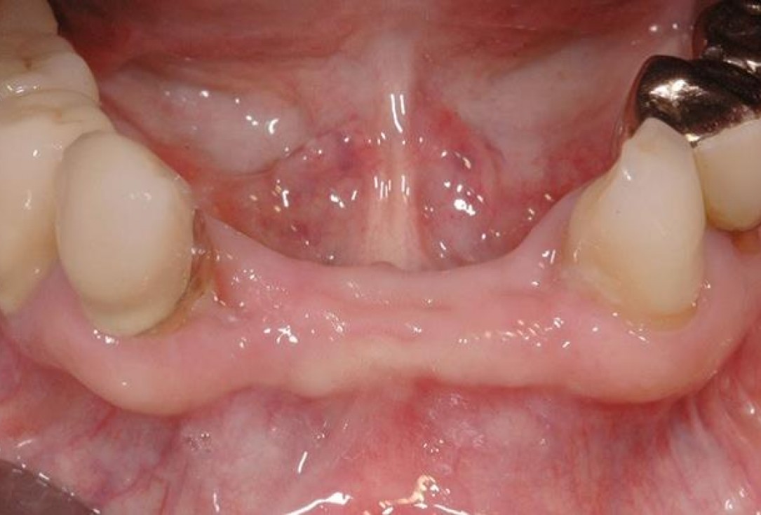
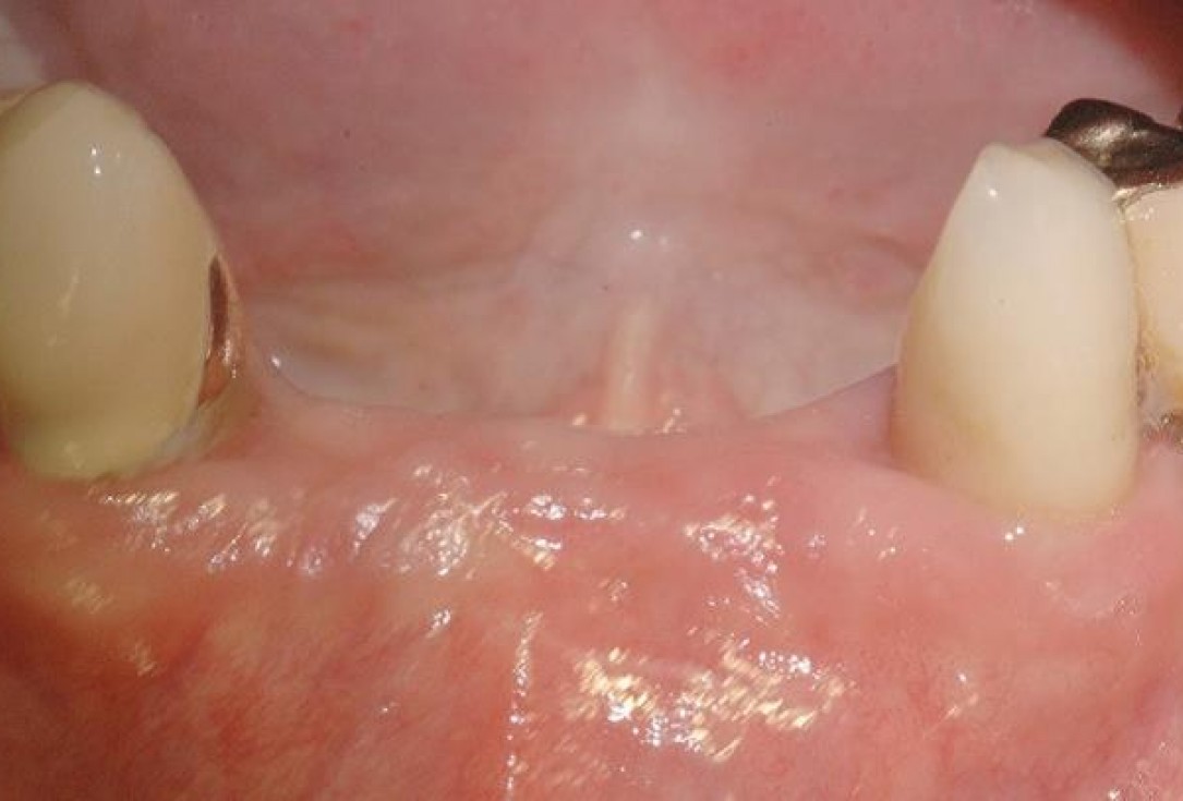
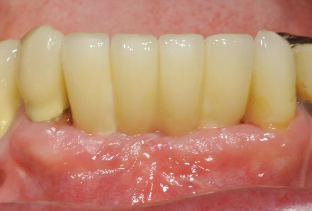
2/15 - 3 months after extraction visible bone and soft tissue loss in regio 42-32Soft tissue augmentation and GBR with mucoderm® and maxresorb® - Dr. S. Scherg
-
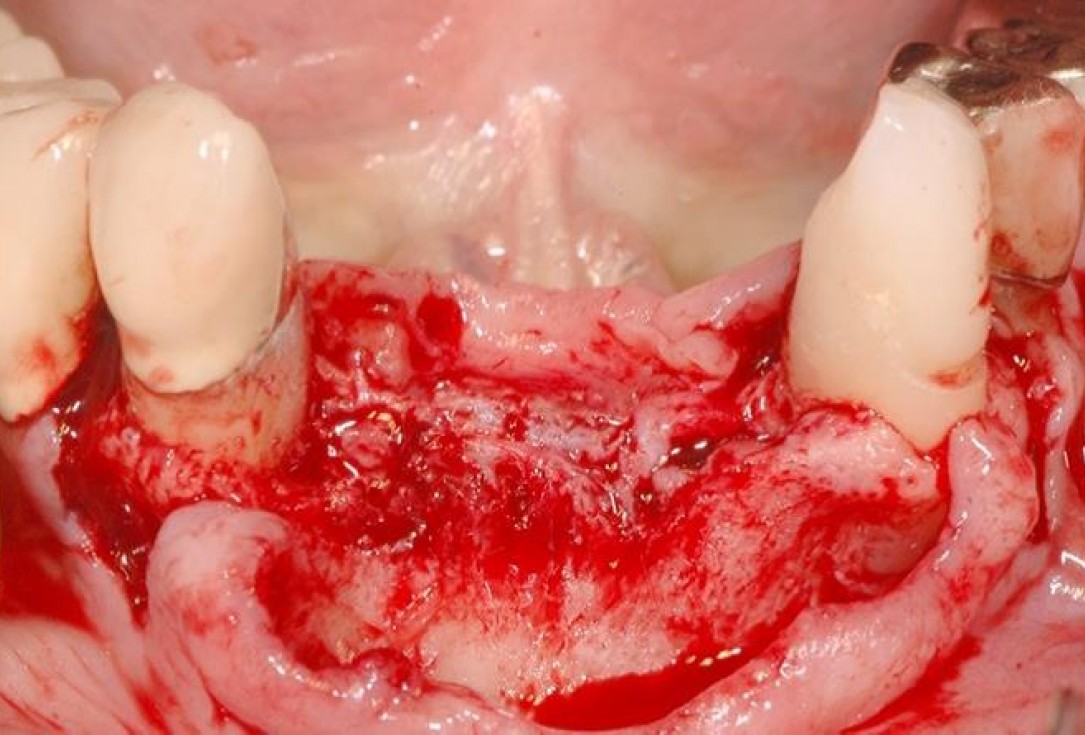
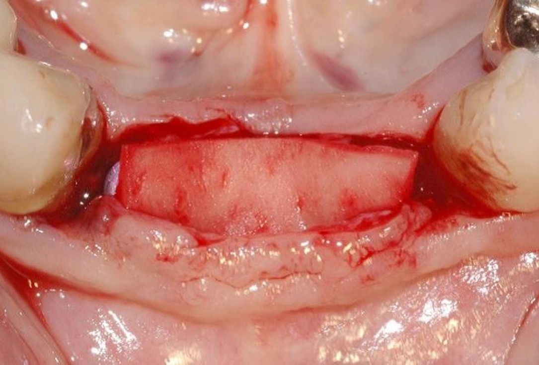
3/15 - Defect dimensions visible after full thickness flap preparationSoft tissue augmentation and GBR with mucoderm® and maxresorb® - Dr. S. Scherg
-
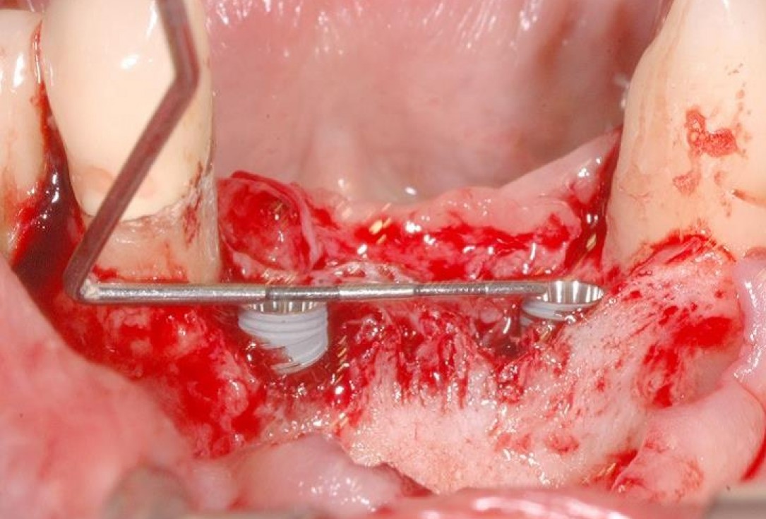
4/15 - Implants placed in the correct positionSoft tissue augmentation and GBR with mucoderm® and maxresorb® - Dr. S. Scherg
-
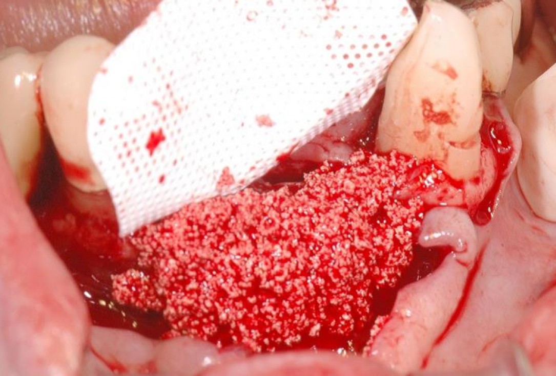
5/15 - Ridge augmentation with maxresorb® and covering with a fixed non-resorbable membraneSoft tissue augmentation and GBR with mucoderm® and maxresorb® - Dr. S. Scherg
-
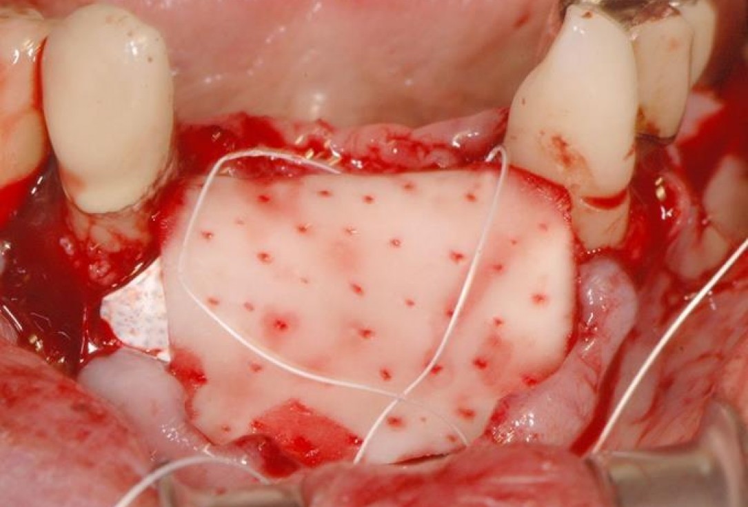
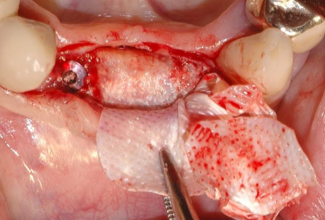
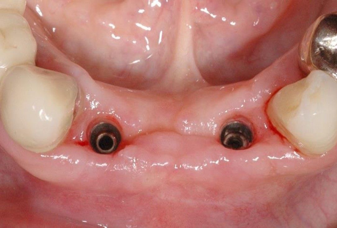
6/15 - Soft tissue augmentation with mucoderm® and fixation with the flapSoft tissue augmentation and GBR with mucoderm® and maxresorb® - Dr. S. Scherg
-
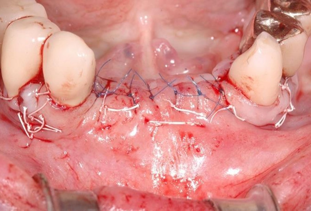
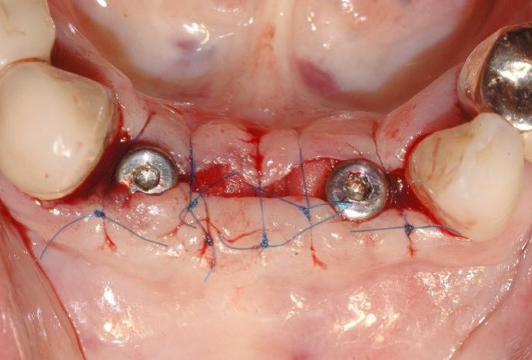
7/15 - Tension-free wound closure with different suturesSoft tissue augmentation and GBR with mucoderm® and maxresorb® - Dr. S. Scherg
-
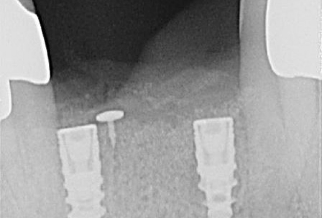
8/15 - X-ray control shows implants and fixation pins directly after surgerySoft tissue augmentation and GBR with mucoderm® and maxresorb® - Dr. S. Scherg
-
9/15 - Uneventful wound healing at 4 monthsSoft tissue augmentation and GBR with mucoderm® and maxresorb® - Dr. S. Scherg
-
10/15 - Re-entry and removal of the non-resorbable membraneSoft tissue augmentation and GBR with mucoderm® and maxresorb® - Dr. S. Scherg
-
11/15 - Further soft tissue augmentation with mucoderm®Soft tissue augmentation and GBR with mucoderm® and maxresorb® - Dr. S. Scherg
-
12/15 - Tension-free wound closure with healing abutmentsSoft tissue augmentation and GBR with mucoderm® and maxresorb® - Dr. S. Scherg
-
13/15 - Healing 4 weeks after re-entry and visible augmentation of the buccal zoneSoft tissue augmentation and GBR with mucoderm® and maxresorb® - Dr. S. Scherg
-
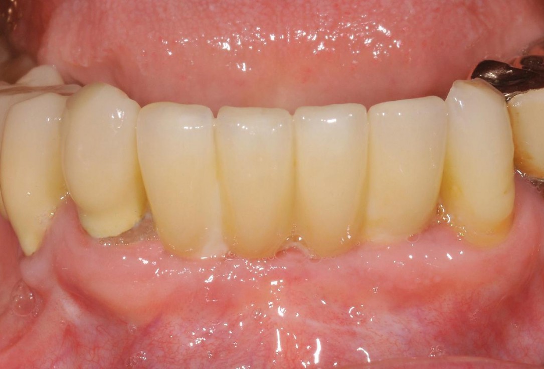
14/15 - Clincal outcome with fixed prosthesisSoft tissue augmentation and GBR with mucoderm® and maxresorb® - Dr. S. Scherg
-
15/15 - Clinical outcome at 24 months with stable soft tissueSoft tissue augmentation and GBR with mucoderm® and maxresorb® - Dr. S. Scherg

Initial Orthopantomograph X-Ray

Pre-operative x-ray

X-ray control before tooth extraction

DVT image demonstrating horizontal and vertical amount of bone available

Surgical presentation of the alveolar ridge with reduced amount of horizontal bone available

DVT control after sinusitis surgery, residual bone height 1 mm

Clinical situation before extraction

DVT control after sinusitis surgery, residual bone height 1 mm

DVT image showing the reduced amount of bone available in the area of the mental foramen

X-ray shows a 3-dimensional periondontal defect

Initial situation: Inflammated tooth #12
