Bone augmentation in aesthetic zone with maxgraft® bonering - Dr. A. Patel
-
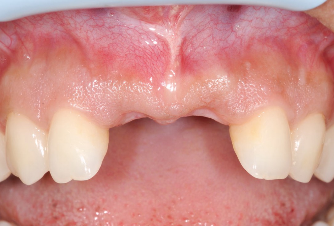
1/26 - Initially bridge retained incisorsBone augmentation in aesthetic zone with maxgraft® bonering - Dr. A. Patel
-
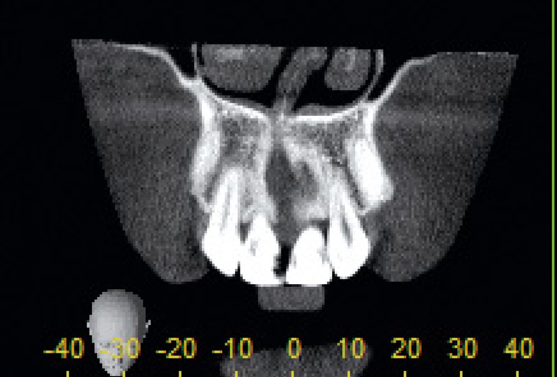
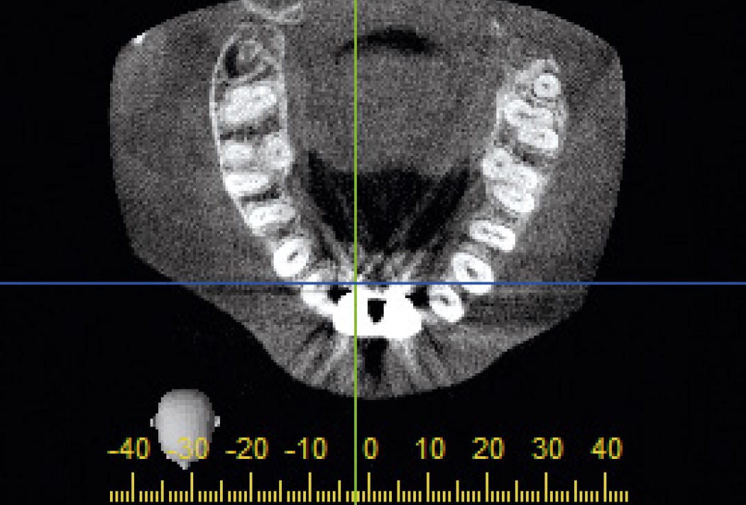
2/26 - CT scan reveals major bone loss in frontal maxillaBone augmentation in aesthetic zone with maxgraft® bonering - Dr. A. Patel
-
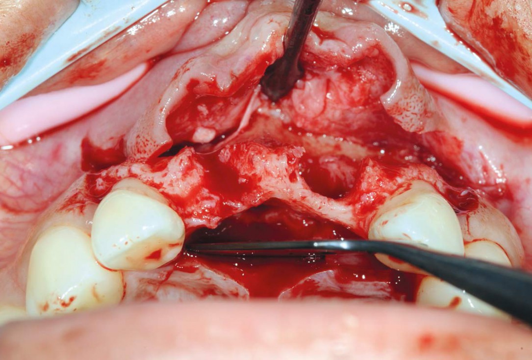
3/26 - Big bone defect visible after opening the flapBone augmentation in aesthetic zone with maxgraft® bonering - Dr. A. Patel
-
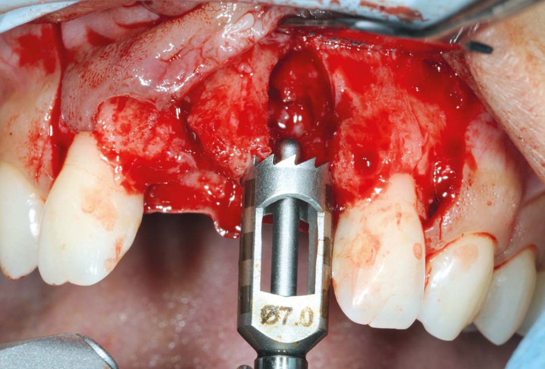
4/26 - Determine the defect size with the trephineBone augmentation in aesthetic zone with maxgraft® bonering - Dr. A. Patel
-
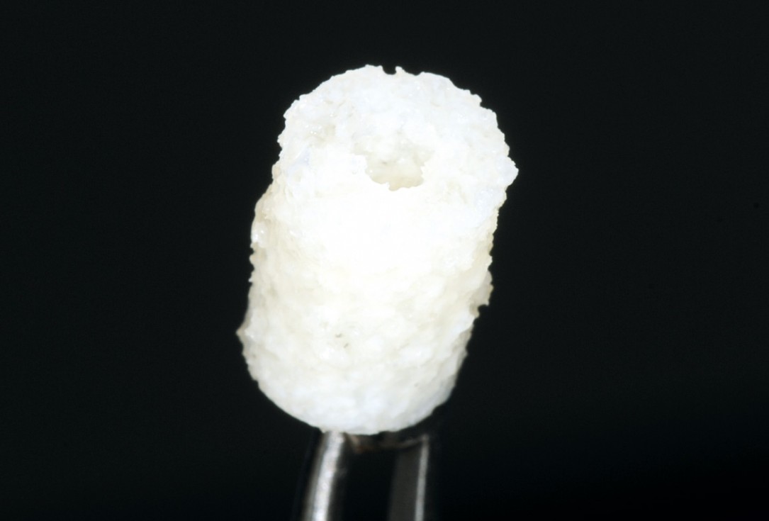
5/26 - Hydrated maxgraft® bonering 7 mmBone augmentation in aesthetic zone with maxgraft® bonering - Dr. A. Patel
-
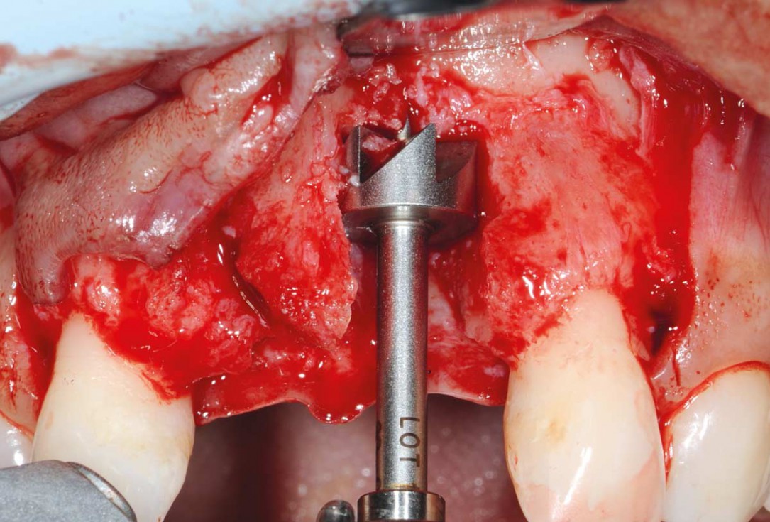
6/26 - Preparing the site for maxgraft® boneringBone augmentation in aesthetic zone with maxgraft® bonering - Dr. A. Patel
-
7/26 - Paving the surface of the recipient site to create press-fitting of the ringBone augmentation in aesthetic zone with maxgraft® bonering - Dr. A. Patel
-
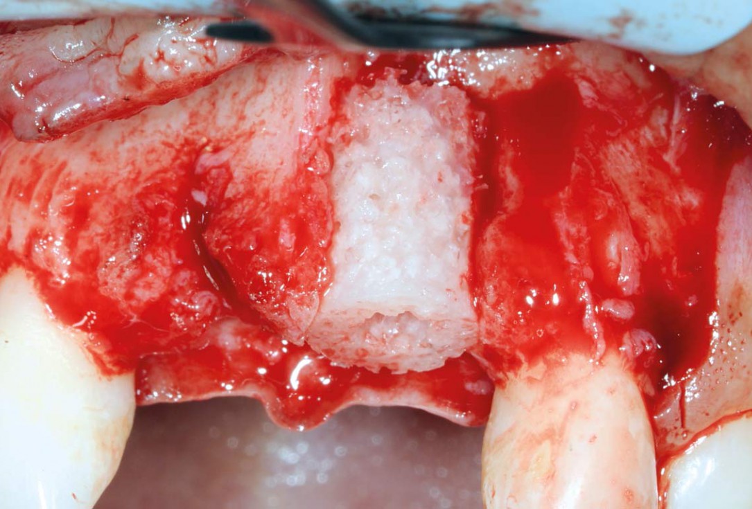
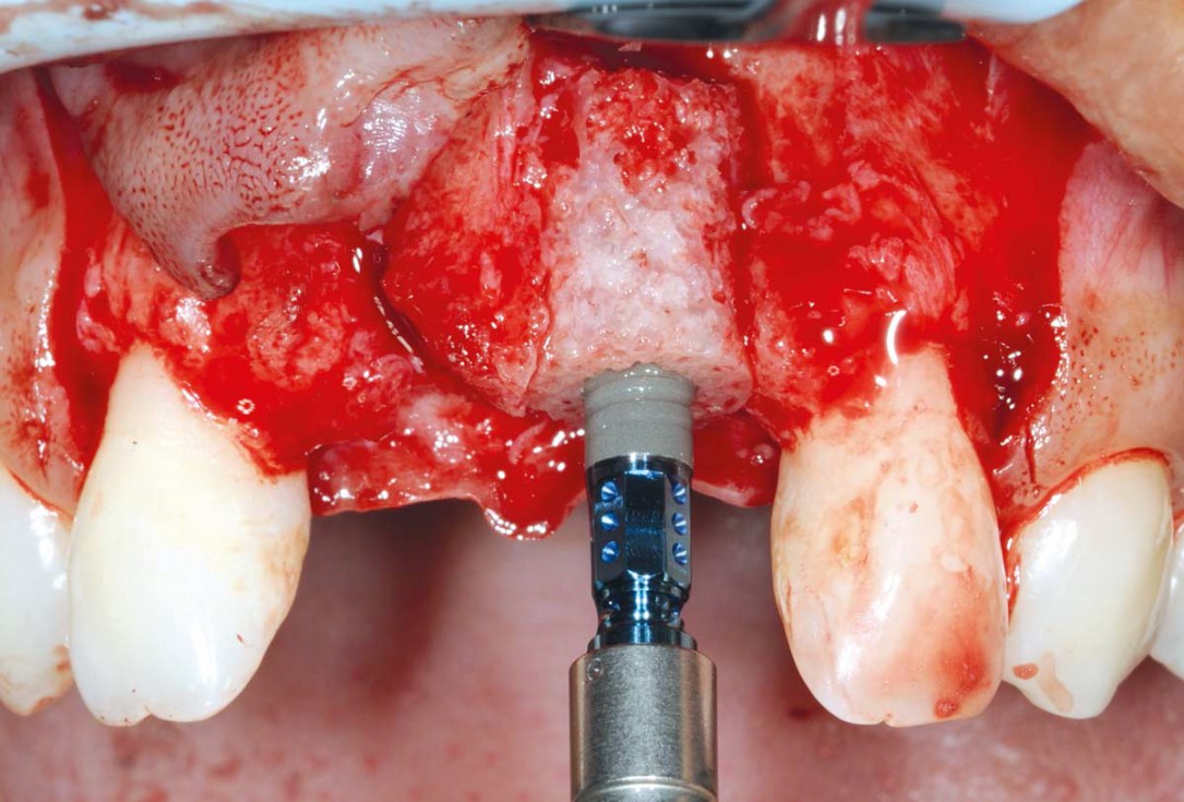
8/26 - Placement of maxgraft® bonering 10 mm heightBone augmentation in aesthetic zone with maxgraft® bonering - Dr. A. Patel
-
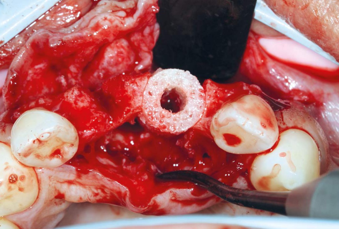
9/26 - Occlusal view after placing the bone graft confirms ideal positioningBone augmentation in aesthetic zone with maxgraft® bonering - Dr. A. Patel
-
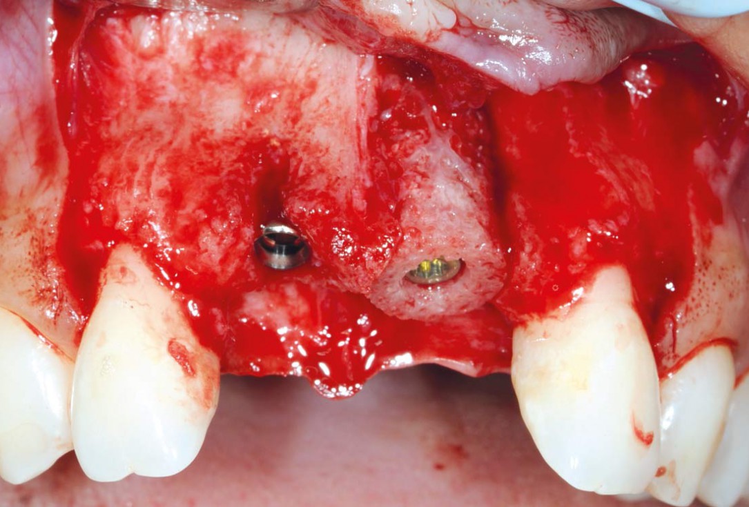
10/26 - Fixation of maxgraft® bonering with a Straumann® SLActive Bone Level Tapered ImplantBone augmentation in aesthetic zone with maxgraft® bonering - Dr. A. Patel
-
11/26 - Implantation in region 11Bone augmentation in aesthetic zone with maxgraft® bonering - Dr. A. Patel
-
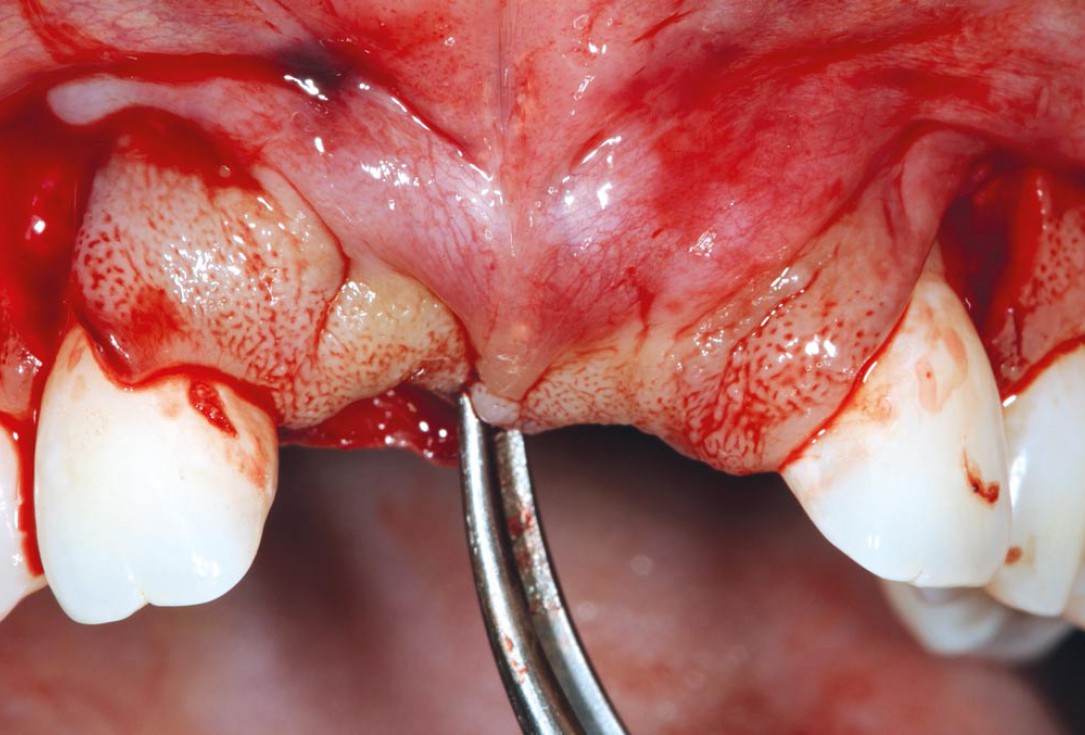
12/26 - Mobilizing the flapBone augmentation in aesthetic zone with maxgraft® bonering - Dr. A. Patel
-
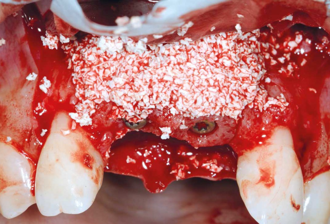
13/26 - Covering the site with cerabone® to prevent resorptionBone augmentation in aesthetic zone with maxgraft® bonering - Dr. A. Patel
-
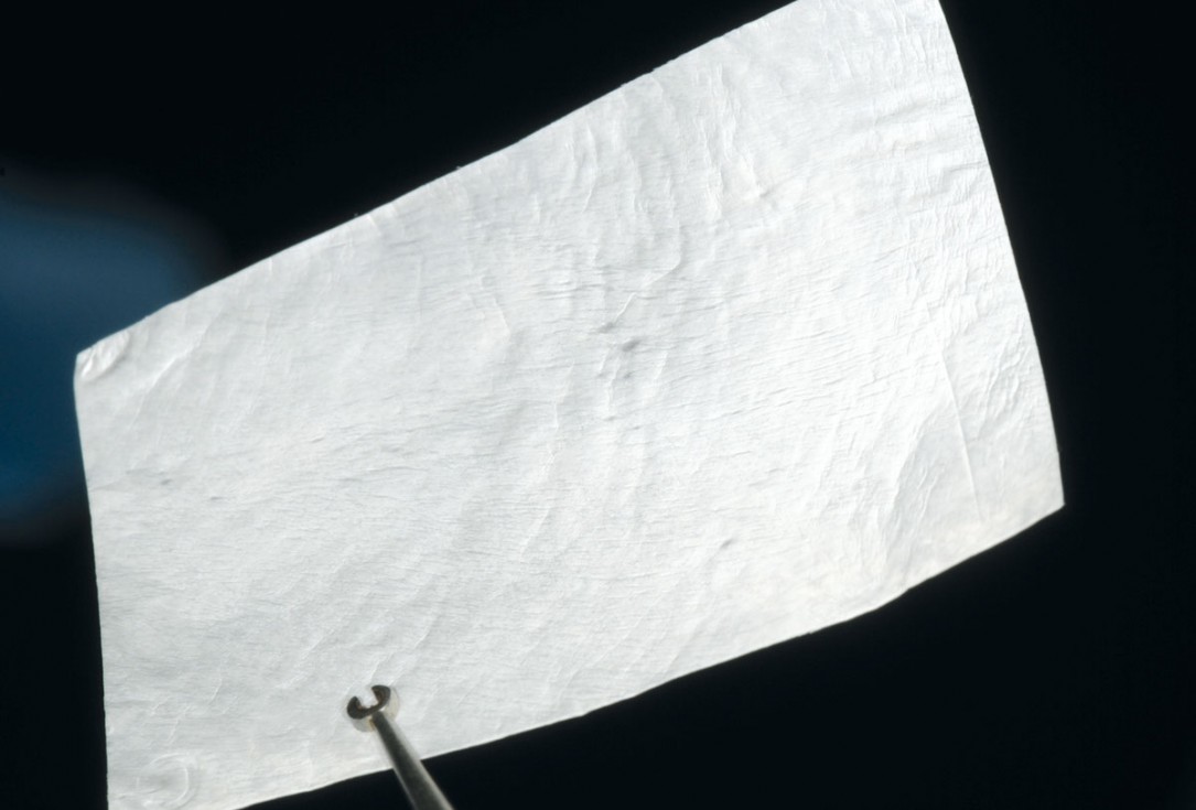
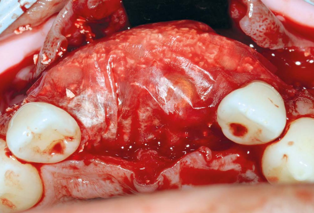
14/26 - Jason® membraneBone augmentation in aesthetic zone with maxgraft® bonering - Dr. A. Patel
-
15/26 - Adding more cerabone® after attaching the Jason® membraneBone augmentation in aesthetic zone with maxgraft® bonering - Dr. A. Patel
-
16/26 - Bone volume gained buccally and site covered with Jason® membraneBone augmentation in aesthetic zone with maxgraft® bonering - Dr. A. Patel
-
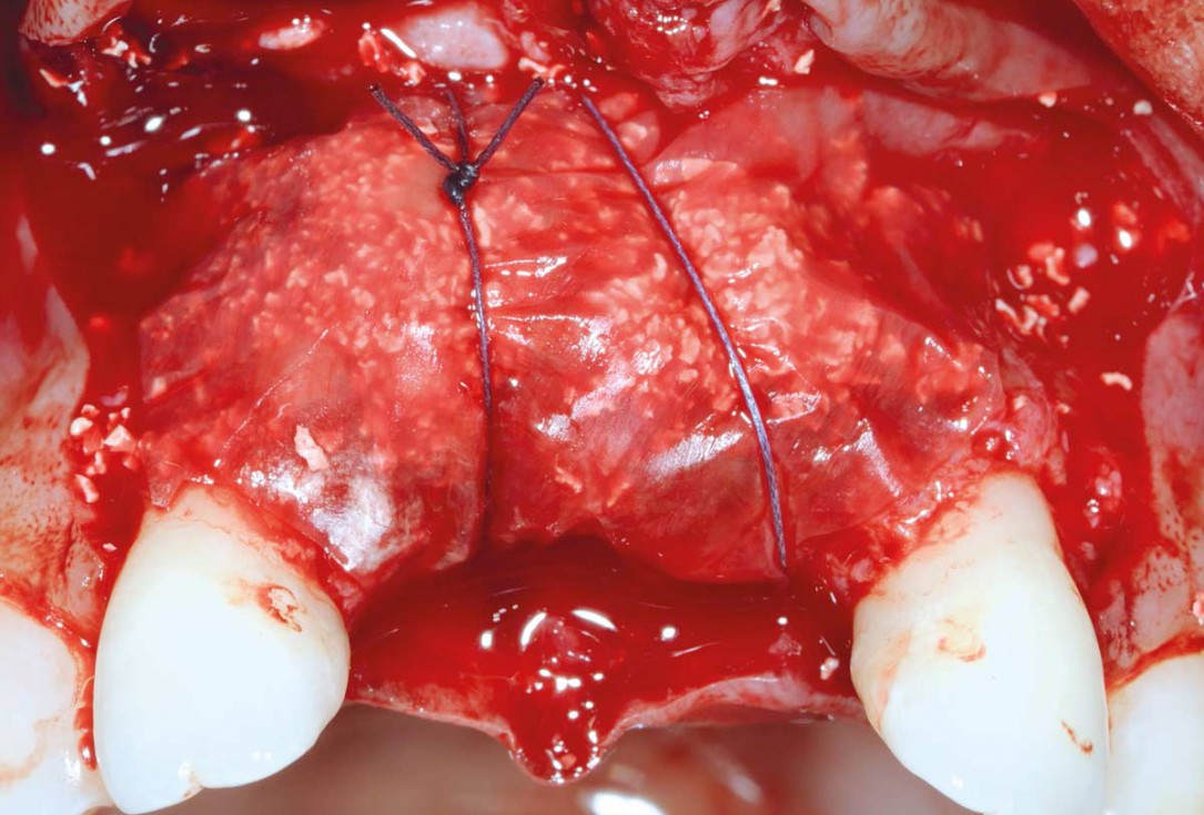
17/26 - Mattress sutures to stabilize the graftBone augmentation in aesthetic zone with maxgraft® bonering - Dr. A. Patel
-
18/26 - Sutured free of tension with vycrilBone augmentation in aesthetic zone with maxgraft® bonering - Dr. A. Patel
-
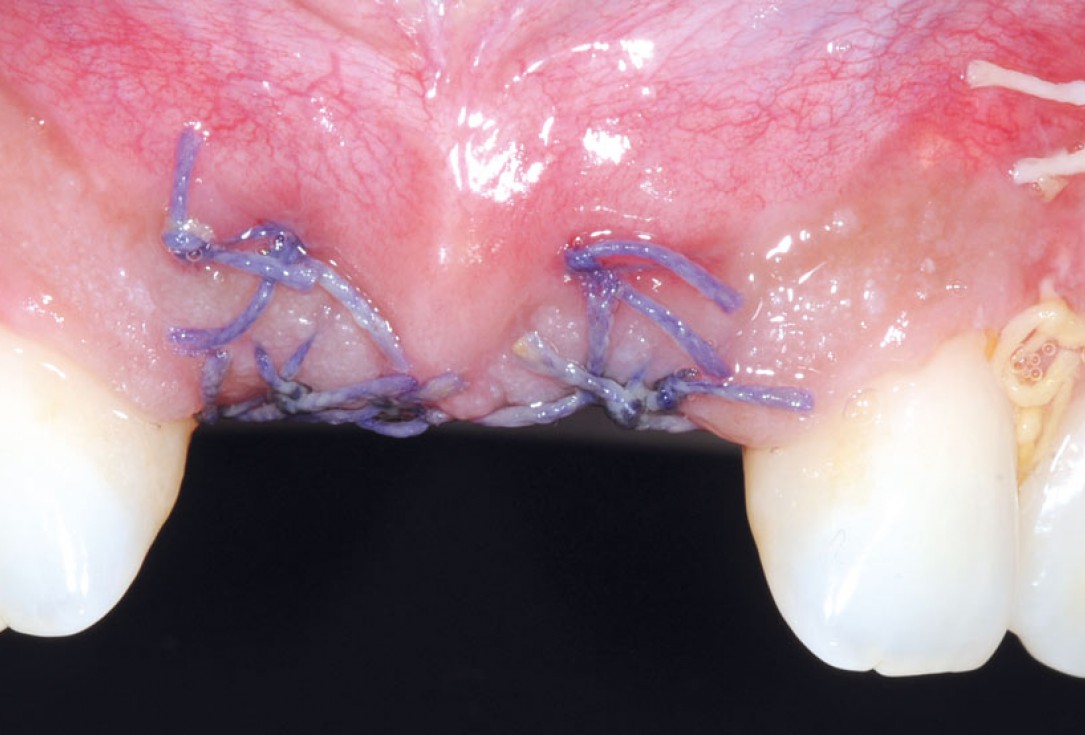
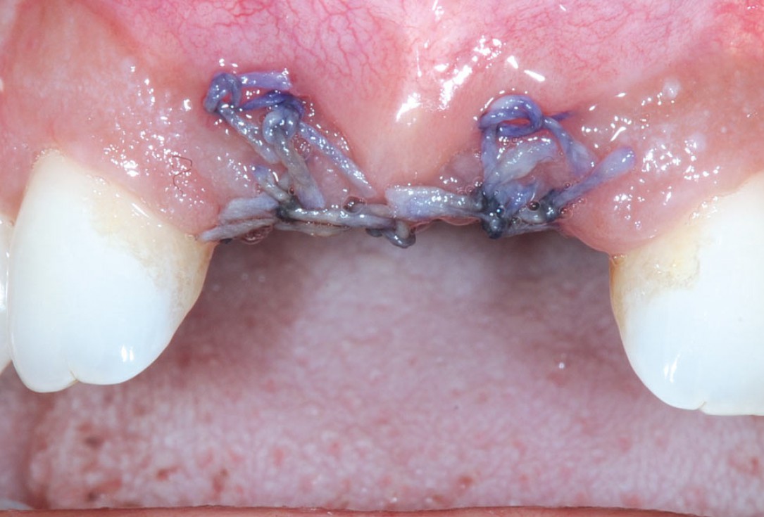
19/26 - 3 weeks post-op: eventless healingBone augmentation in aesthetic zone with maxgraft® bonering - Dr. A. Patel
-
20/26 - 6 weeks post-op: sutures were removedBone augmentation in aesthetic zone with maxgraft® bonering - Dr. A. Patel
-
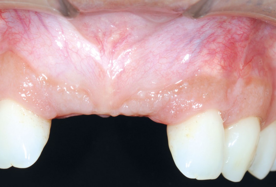
21/26 - 6 months after surgery: healthy soft tissuesBone augmentation in aesthetic zone with maxgraft® bonering - Dr. A. Patel
-
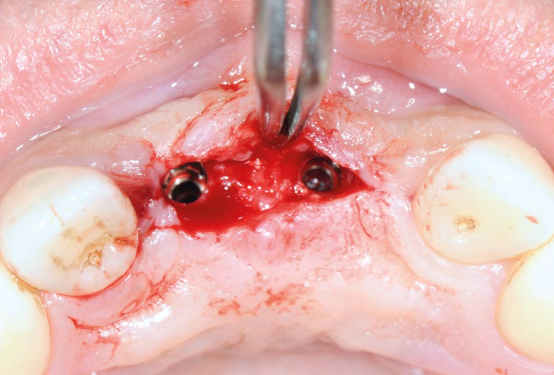
22/26 - Uncovering the implants 6 months after surgeryBone augmentation in aesthetic zone with maxgraft® bonering - Dr. A. Patel
-
23/26 - Prosthetic rehabilitationBone augmentation in aesthetic zone with maxgraft® bonering - Dr. A. Patel
-
24/26 - CT scan after implants been restoredBone augmentation in aesthetic zone with maxgraft® bonering - Dr. A. Patel
-
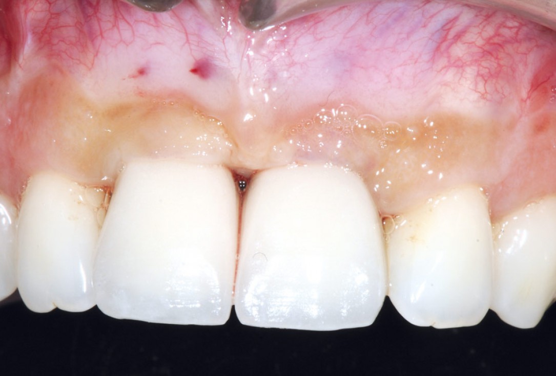
25/26 - Final crowns immediatly after restorationBone augmentation in aesthetic zone with maxgraft® bonering - Dr. A. Patel
-
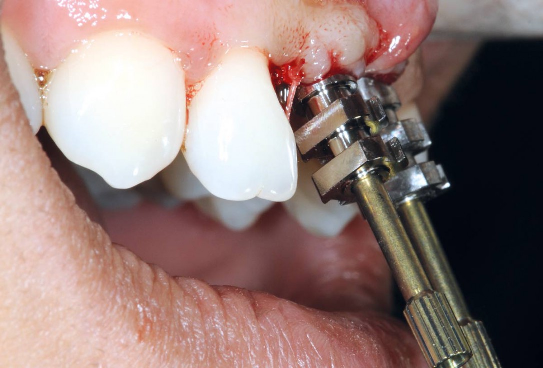
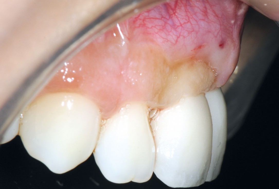
26/26 - Lateral view confirms bone volume gainBone augmentation in aesthetic zone with maxgraft® bonering - Dr. A. Patel

Vertical augmentation: Preparation of ring bed in atrophic mandibula (third quadrant)

Initial situation pre-op: Central incisors with mobility 3

Severe periimplantitis at tooth 15 with bone loss up to 1/3 of the implant

Initial situation: X-ray scan reveals eggshell thin sinus floor (1-3 mm) on both sites of the maxilla; green areas indicate the planned maxgraft® bonerings and red areas the planned implants

Initial presentation of failing post retained crown with previous history of failed apicectomies and amalgam tattooing and scar tissue

Pilot drilling in the recipient site

X-ray scan of clinical situation

Initial situation 57-year old female patient. X-ray scan reveals severe bone loss due to inflammation in region 13. Treatment plan was extraction of teeth 13 and 14 and augmentation after healing.

X-ray scan: initial situation loss of two wall bony defect with loss of buccal and lingual lamella

Initial situation: single tooth gap in regio 22

Bone defect in the aesthetic zone.

Initial situation: missing incisor with loss of buccal wall

Planning the surgery with CoDiagnostix® for Straumann® Guided Surgery

X-ray scan reveals initial situation with maxillary bone height in regio 15 of 1.5 mm

Initial situation: bone loss due to lack of physical load of bridge retained region 11
