Non-contained intrabony defect treated with the modified papilla preservation flap in conjunction with cerabone® and Straumann® Emdogain® - Dr. J. Tunkel
-
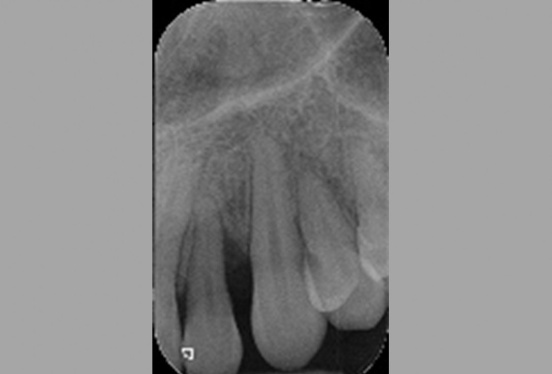
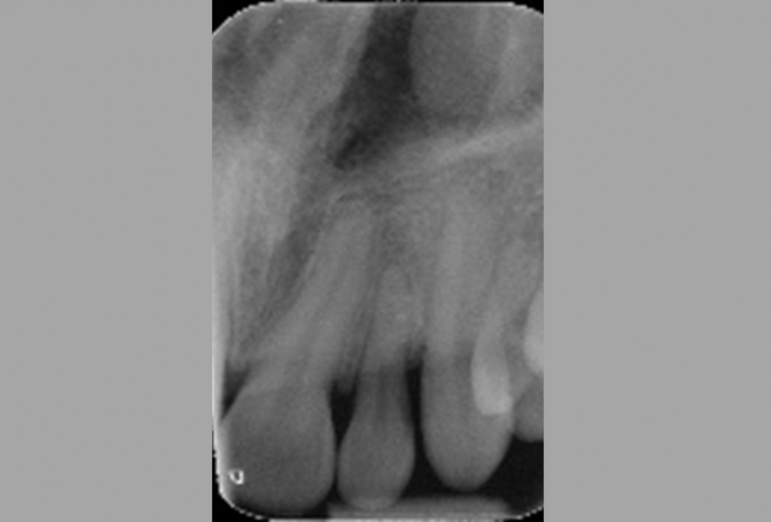
01/11 - Pre-operative radiographic view. Intrabony defect on the distal aspect of the lateral incisor.Non-contained intrabony defect treated with the modified papilla preservation flap in conjunction with cerabone® and Straumann® Emdogain® - Dr. J. Tunkel
-
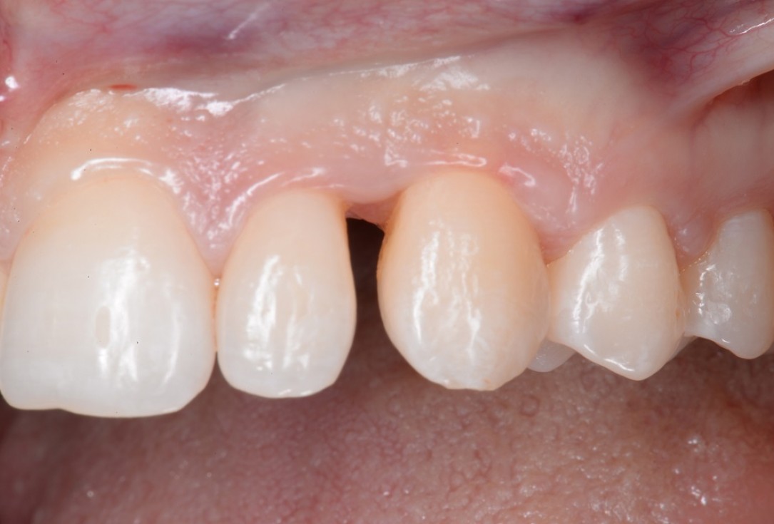
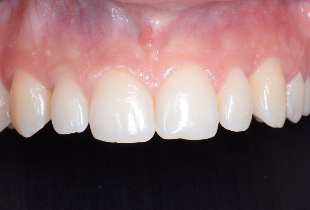
02/11 - Pre-clinical situation.Non-contained intrabony defect treated with the modified papilla preservation flap in conjunction with cerabone® and Straumann® Emdogain® - Dr. J. Tunkel
-
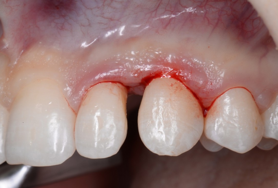
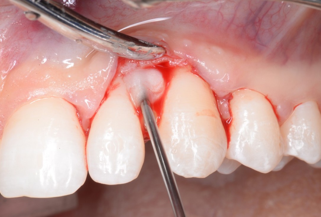
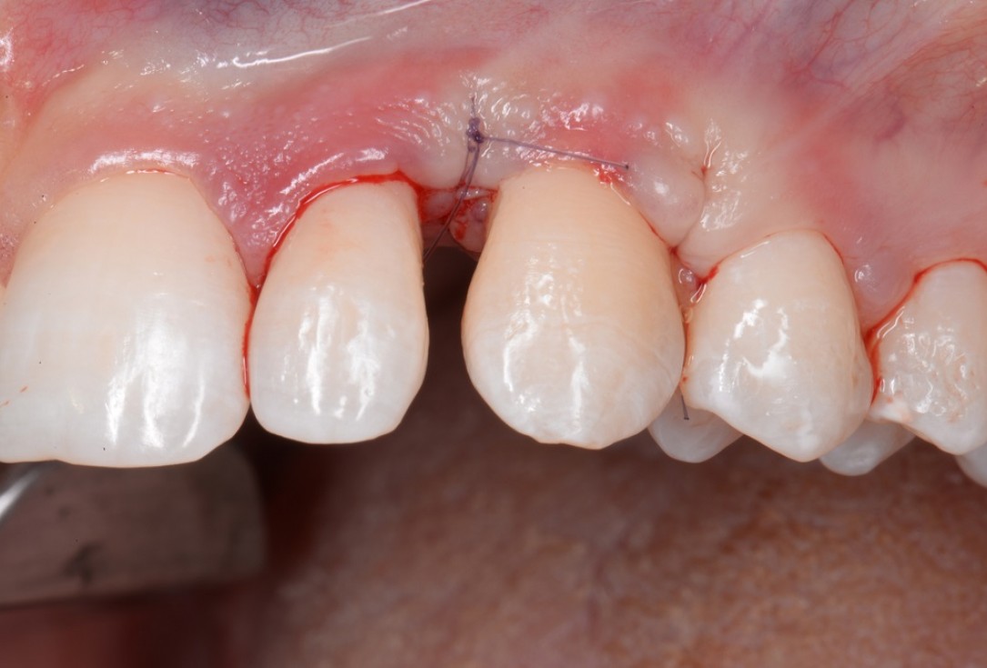
03/11 - Incisions according to the modified papilla preservation flap (MPPF) technique (Cortellini et al. J Periodontol. 1995).Non-contained intrabony defect treated with the modified papilla preservation flap in conjunction with cerabone® and Straumann® Emdogain® - Dr. J. Tunkel
-
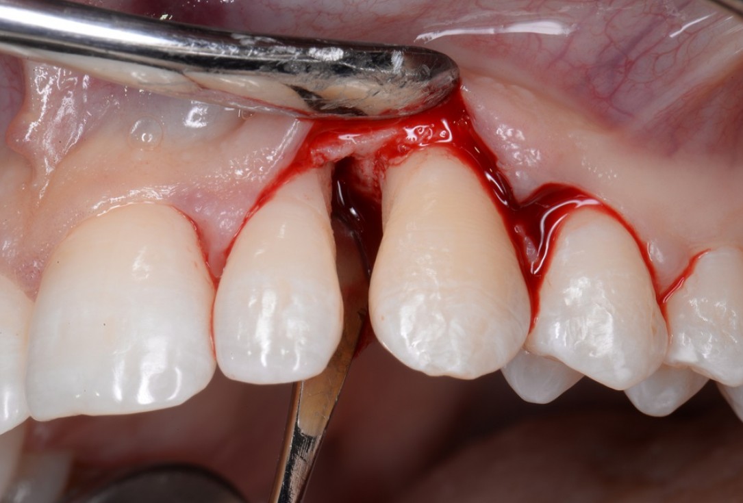
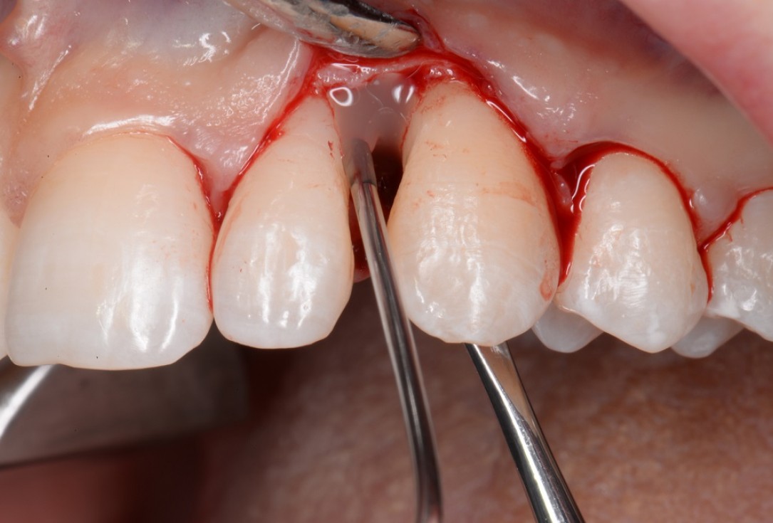
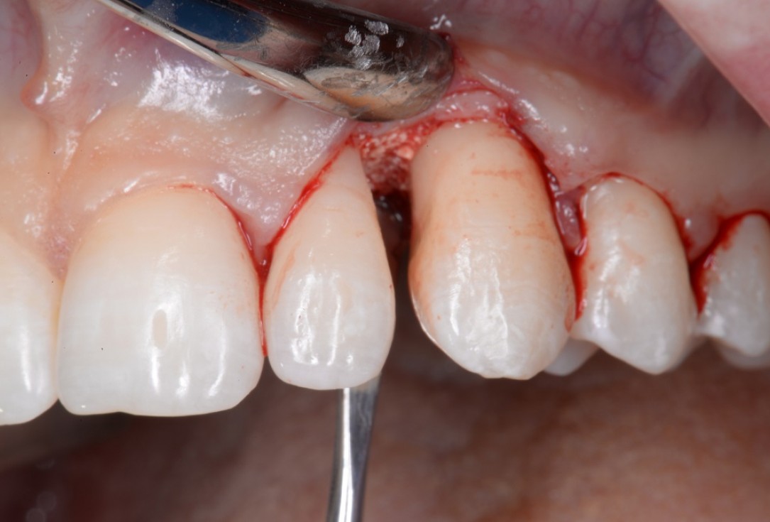
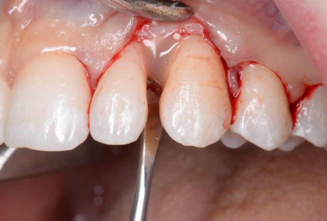
04/11 - Flap elevation according to the MPPF technique (Cortellini et al. J Periodontol. 1995). The defect presented as mixed 1-, 2- and 3-wall defect.Non-contained intrabony defect treated with the modified papilla preservation flap in conjunction with cerabone® and Straumann® Emdogain® - Dr. J. Tunkel
-
05/11 - Application of Straumann® PrefGel® to the root surface.Non-contained intrabony defect treated with the modified papilla preservation flap in conjunction with cerabone® and Straumann® Emdogain® - Dr. J. Tunkel
-
06/11 - Application of Straumann® Emdogain® to the root surface.Non-contained intrabony defect treated with the modified papilla preservation flap in conjunction with cerabone® and Straumann® Emdogain® - Dr. J. Tunkel
-
07/11 - Application of cerabone® granules.Non-contained intrabony defect treated with the modified papilla preservation flap in conjunction with cerabone® and Straumann® Emdogain® - Dr. J. Tunkel
-
08/11 - Application of Straumann® Emdogain® on the bone graft directly before flap closure.Non-contained intrabony defect treated with the modified papilla preservation flap in conjunction with cerabone® and Straumann® Emdogain® - Dr. J. Tunkel
-
09/11 - Flap closure.Non-contained intrabony defect treated with the modified papilla preservation flap in conjunction with cerabone® and Straumann® Emdogain® - Dr. J. Tunkel
-
10/11 - Clinical situation 8 months post-operative.Non-contained intrabony defect treated with the modified papilla preservation flap in conjunction with cerabone® and Straumann® Emdogain® - Dr. J. Tunkel
-
11/11 - Radiographic view 8 months post-operative.Non-contained intrabony defect treated with the modified papilla preservation flap in conjunction with cerabone® and Straumann® Emdogain® - Dr. J. Tunkel
![01/23 GBR of the edentulous maxillary ridge using permamem®, cerabone® and autologous bone chips - Dr. V. Kalenchuk](https://www.indication-matrix.de/sites/default/files/styles/gridimagesbreakpoints_theme_botiss_mobile_1x/public/caseimages/botiss-cerabone-permamem-gbr-edentulous-ridge-kalenchuk-01.jpg?itok=97VwjfQZ×tamp=1584977530)
Initial clinical situation. Atrophic maxillary ridge.
![01/32 Regeneration of a 9 mm vertical bone defect with cerabone®, autologous bone and S-PRF – Dr. A. Eslava](https://www.indication-matrix.de/sites/default/files/styles/gridimagesbreakpoints_theme_botiss_mobile_1x/public/caseimages/1-initial-x-ray-implants.jpg?itok=lBN0dalH×tamp=1606145751)
Initial x-ray showing bone loss around implants placed 5 years ago in another dental clinic
![1/18 cerabone® and mucoderm® for immediacy in esthetic zone -Dr. M Motta](https://www.indication-matrix.de/sites/default/files/styles/gridimagesbreakpoints_theme_botiss_mobile_1x/public/caseimages/casos-botiss.015.jpg?itok=sdIJ9Lov×tamp=1565947808)
Initial view of the case. Discoloration of 1.1 and mild class I gingival recession
![01/11 Ridge augmentation using cerabone®, Jason® membrane and Straumann® Emdogain® - Dr. S. Pelekanos](https://www.indication-matrix.de/sites/default/files/styles/gridimagesbreakpoints_theme_botiss_mobile_1x/public/caseimages/botiss-cerabone-jason-membrane-emdogain-ridge-augmentation-pelekanos-1_0.jpg?itok=MNP_Qtan×tamp=1531381370)
Situation after tooth removal.
![01/19 GBR with cerabone® and Jason® membrane in the front tooth region - Dr. H. Maghaireh](https://www.indication-matrix.de/sites/default/files/styles/gridimagesbreakpoints_theme_botiss_mobile_1x/public/caseimages/botiss_cerabone_jason_maghaireh_1.jpg?itok=o9sKIZMB×tamp=1495799300)
Initial clinical situation with gum recession and labial bone loss eight weeks following tooth extraction
![01/07 botiss cerabone® & Jason® membrane for horizontal augmentation - Clinical case by Dr. M. Steigmann](https://www.indication-matrix.de/sites/default/files/styles/gridimagesbreakpoints_theme_botiss_mobile_1x/public/caseimages/botiss-cerabone-jason-membrane-gbr-horizontal-augmentatation-steigmann-1.jpg?itok=Bdu6nboX×tamp=1424873438)
Three implants placed in a narrow posterior mandible
![01/12 Two-wall intrabony defect treated using cerabone® and Straumann® Emdogain® - Rakasevic & Jankovic](https://www.indication-matrix.de/sites/default/files/styles/gridimagesbreakpoints_theme_botiss_mobile_1x/public/caseimages/bild1.jpg?itok=mg3zlt8C×tamp=1462803612)
Pre-operative clinical situation.
![01/13 botiss cerabone®, Jason® membrane & collagen fleece for two-stage sinus lift - Clinical case by Dr. D. Jelušić](https://www.indication-matrix.de/sites/default/files/styles/gridimagesbreakpoints_theme_botiss_mobile_1x/public/caseimages/botiss-cerabone-jason-membrane-fleece-sinus-lift-two-stage-jelusic-2.jpg?itok=PJKkUW0Q×tamp=1424880562)
Preoperative clinical situation
![01/15 One-stage sinus lift with cerabone® and Jason® membrane - Dr. Damir Jelušić](https://www.indication-matrix.de/sites/default/files/styles/gridimagesbreakpoints_theme_botiss_mobile_1x/public/caseimages/1-sinus_lift-jelusic.jpg?itok=M6HlPcLx×tamp=1412870443)
Pre-operative OPG
![01/10 Vertical and horizontal GBR using permamem®, cerabone® and autologous bone - Dr. D. B. Hangyasi](https://www.indication-matrix.de/sites/default/files/styles/gridimagesbreakpoints_theme_botiss_mobile_1x/public/caseimages/botiss-cerabone-permamem-vertical-horizontal-gbr-hangyasi-01.jpg?itok=QjxvOXo7×tamp=1589455377)
Initial clinical situation.
![01/13 Vertical bone augmentation using tent pole screw-technique - Prof. Dr. S Fickl](https://www.indication-matrix.de/sites/default/files/styles/gridimagesbreakpoints_theme_botiss_mobile_1x/public/caseimages/botiss-cerabone-permamem-vertical-ridge-augmentation-fickl-01.jpg?itok=NGCPzoD_×tamp=1584799373)
Pre-surgical situation.
![1/27 GBR and soft tissue augmentation with cerabone® and mucoderm® - H. Maghaireh & V. Ivancheva](https://www.indication-matrix.de/sites/default/files/styles/gridimagesbreakpoints_theme_botiss_mobile_1x/public/caseimages/1_11.jpg?itok=gP97Ldm8×tamp=1557222058)
Initial situation: missing teeth #11 & 12 and badly broken #21 root
![01/10](https://www.indication-matrix.de/sites/default/files/styles/gridimagesbreakpoints_theme_botiss_mobile_1x/public/caseimages/botiss-collprotect-cerabone-straumann-emdogain-intraossaerer-defekt-schwaar1.jpg?itok=upWS6HES×tamp=1525702321)
Pre-operative OPG shows deep vertical intrabony defects on the distal aspects of teeth 13 and 14.
![01/18 botiss cerabone® & Jason® membrane for GBR - Clinical case by Prof. Dr. Dr. D. Rothamel](https://www.indication-matrix.de/sites/default/files/styles/gridimagesbreakpoints_theme_botiss_mobile_1x/public/caseimages/botiss-cerabone-jason-membrane-gbr-rothamel-1.jpg?itok=ATsIE-WB×tamp=1424871475)
Instable bridge situation with abscess formation at tooth #15 after apicoectomy
![01/12 Horizontal ridge augmentation using cerabone® block, permamem® and Jason® membrane - Dr. P. Lázaro](https://www.indication-matrix.de/sites/default/files/styles/gridimagesbreakpoints_theme_botiss_mobile_1x/public/caseimages/botiss-horizontal-gbr-permamem-cerabone-jason-membrane-lazaro-1.jpg?itok=2yAK7SFs×tamp=1588251406)
Initial clinical situation.
![01/11 botiss cerabone® & Jason® membrane for GBR - Clinical case by Dr. S. Kovalevsky](https://www.indication-matrix.de/sites/default/files/styles/gridimagesbreakpoints_theme_botiss_mobile_1x/public/caseimages/botiss-cerabone-jason-membrane-gbr-horizontal-augmentatation-kovalevsky-1.jpg?itok=kNjsWze4×tamp=1424876064)
Implant insertion in atrophic alveolar ridge
![01/10 botiss cerabone® & collagen fleece for immediate implantation - Clinical case by Dr. D. Jelušić](https://www.indication-matrix.de/sites/default/files/styles/gridimagesbreakpoints_theme_botiss_mobile_1x/public/caseimages/botiss-cerabone-jason-fleece-immediate-implantation-jelusic-1.jpg?itok=tyKFOrLA×tamp=1424861391)
Clinical situation before extraction and implantation
![01/06](https://www.indication-matrix.de/sites/default/files/styles/gridimagesbreakpoints_theme_botiss_mobile_1x/public/caseimages/bild1_3_new.jpg?itok=fFRO4F24×tamp=1572522845)
Pre-operative radiographic view.
![1/15 Socket preservation using cerabone®, Jason® membrane and permamem® - Dr. P. Di Capua](https://www.indication-matrix.de/sites/default/files/styles/gridimagesbreakpoints_theme_botiss_mobile_1x/public/caseimages/botiss-permamem-socket-preservation-di-capua-1.jpg?itok=QiAadTWG×tamp=1502716540)
Situation after tooth extraction.
![01/17 Socket preservation using cerabone® and Straumann® Emdogain® - Dr. S. Pelekanos](https://www.indication-matrix.de/sites/default/files/styles/gridimagesbreakpoints_theme_botiss_mobile_1x/public/caseimages/botiss-cerabone-emdogain-socket-preservation-pelekanos-0.jpg?itok=eE540ojG×tamp=1531378890)
Pre-operative X-ray. Hopless tooth 21.
![01/16 Two-stage sinus floor elevation and horizontal GBR using cerabone®, autologous bone and Jason® membrane - Dr. M. Blašković](https://www.indication-matrix.de/sites/default/files/styles/gridimagesbreakpoints_theme_botiss_mobile_1x/public/caseimages/botiss-cerabone-autogenous-bone-jason_membrane-sfe-gbr-blaskovic-1.jpg?itok=zZq4UMwk×tamp=1521368066)
Pre-surgical situation. Teeth 26 and 27 missing.
![01/12 botiss cerabone® & Jason® membrane for treatment of dehiscence defect - Clinical case by Dr. M. Steigmann](https://www.indication-matrix.de/sites/default/files/styles/gridimagesbreakpoints_theme_botiss_mobile_1x/public/caseimages/f104_steigmann_cerabone-dehiscence.jpg?itok=TQ14OKOA×tamp=1568103962)
Extraction of tooth 21 after endodontic treatment
![01/11 Intrabony defect treated using collprotect® membrane & cerabone® (1) - Cosgarea & Sculean](https://www.indication-matrix.de/sites/default/files/styles/gridimagesbreakpoints_theme_botiss_mobile_1x/public/caseimages/1_0.jpg?itok=qc6wc5t6×tamp=1462786032)
Pre-surgical probing reveals a deep intrabony defect on the distal aspect of the upper canine.
![01/15 botiss cerabone® & Jason® membrane for block augmentation with autologous bone blocks - clinical case by Dr. S. Stavar](https://www.indication-matrix.de/sites/default/files/styles/gridimagesbreakpoints_theme_botiss_mobile_1x/public/caseimages/botiss-cerabone-jason-membrane-block-augmentation-autologous-bone-block-stavar-001a_c.jpg?itok=2DD2cc5N×tamp=1425041275)
Initial clinical situation with single tooth gap in regio 21
![01/08 botiss cerabone® & collprotect® membrane for GBR - Clinical case by Dr. V. Kalenchuk](https://www.indication-matrix.de/sites/default/files/styles/gridimagesbreakpoints_theme_botiss_mobile_1x/public/caseimages/botiss-cerabone-collprotect-membrane-gbr-ridge-augmentatation-kalenchuk-1.jpg?itok=TTdIjl5c×tamp=1424878273)
Clinical situation with narrow alveolar ridge in the lower jaw
![01/10 Ridge preservation and reconstruction of the buccal wall using permamem® and cerabone® - Dr. M. Steigmann](https://www.indication-matrix.de/sites/default/files/styles/gridimagesbreakpoints_theme_botiss_mobile_1x/public/caseimages/botiss-permamem-ridge-preservation-steigmann-1.jpg?itok=N4WCJrCz×tamp=1587400013)
Initial clinical situation showing bone wall defect.