Non-contained intrabony defect treated using cerabone®, collprotect® membrane and Straumann® Emdogain® - Dr. T. Schwaar (1)
-
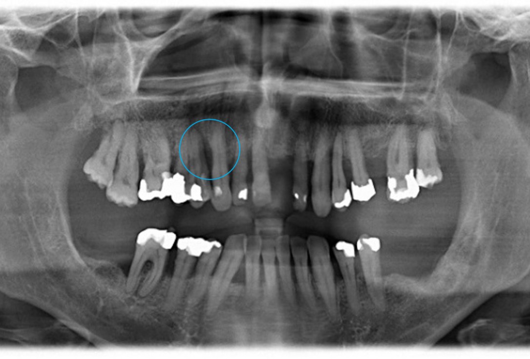
- Pre-operative radiograph. Intrabony defect on the mesial aspect of tooth 14.
 Non-contained intrabony defect treated using cerabone®, collprotect® membrane and Straumann® Emdogain® - Dr. T. Schwaar (1)
 -
02/11 - Pre-operative radiograph. Deep intrabony defect on the mesial aspect of tooth 14.
 Non-contained intrabony defect treated using cerabone®, collprotect® membrane and Straumann® Emdogain® - Dr. T. Schwaar (1)
 -
03/11 - Pre-surgical clinical situation.
 Non-contained intrabony defect treated using cerabone®, collprotect® membrane and Straumann® Emdogain® - Dr. T. Schwaar (1)
 -
04/11 - Probing pocket depth of 12 mm mesially at tooth 14.
 Non-contained intrabony defect treated using cerabone®, collprotect® membrane and Straumann® Emdogain® - Dr. T. Schwaar (1)
 -
05/11 - 1- to 2-wall intrabony defect at the mesio-palatal aspect of tooth 14.
 Non-contained intrabony defect treated using cerabone®, collprotect® membrane and Straumann® Emdogain® - Dr. T. Schwaar (1)
 -
06/11 - After granulation tissue removal, treatment of the root surface with Straumann® Emdogain®.
 Non-contained intrabony defect treated using cerabone®, collprotect® membrane and Straumann® Emdogain® - Dr. T. Schwaar (1)
 -
07/11 - Root surface covered with collprotect® membrane.
 Non-contained intrabony defect treated using cerabone®, collprotect® membrane and Straumann® Emdogain® - Dr. T. Schwaar (1)
 -
08/11 - Defect grafted with small cerabone® granules.
 Non-contained intrabony defect treated using cerabone®, collprotect® membrane and Straumann® Emdogain® - Dr. T. Schwaar (1)
 -
09/11 - cerabone® granules covered with collprotect® membrane.
 Non-contained intrabony defect treated using cerabone®, collprotect® membrane and Straumann® Emdogain® - Dr. T. Schwaar (1)
 -
10/11 - Suturing.
 Non-contained intrabony defect treated using cerabone®, collprotect® membrane and Straumann® Emdogain® - Dr. T. Schwaar (1)
 -
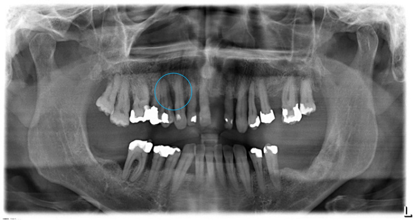
11/11 - Radiographic control 5 months post-operative.
 Non-contained intrabony defect treated using cerabone®, collprotect® membrane and Straumann® Emdogain® - Dr. T. Schwaar (1)


Initial clinical situation. Atrophic maxillary ridge.

Initial x-ray showing bone loss around implants placed 5 years ago in another dental clinic

Initial view of the case. Discoloration of 1.1 and mild class I gingival recession

Situation after tooth removal.


Initial clinical situation with gum recession and labial bone loss eight weeks following tooth extraction

Three implants placed in a narrow posterior mandible

Pre-operative clinical situation.

Clinical situation with narrow alveolar ridge in the lower jaw


Initial clinical situation showing bone wall defect.

Initial clinical situation.

Pre-surgical situation.

Initial situation: missing teeth #11 & 12 and badly broken #21 root

Pre-operative OPG shows deep vertical intrabony defects on the distal aspects of teeth 13 and 14.


Instable bridge situation with abscess formation at tooth #15 after apicoectomy

Initial clinical situation.

Implant insertion in atrophic alveolar ridge

Preoperative clinical situation

Pre-operative OPG


Pre-operative X-ray. Hopless tooth 21.

Pre-surgical situation. Teeth 26 and 27 missing.

Extraction of tooth 21 after endodontic treatment

Pre-surgical probing reveals a deep intrabony defect on the distal aspect of the upper canine.

Initial clinical situation with single tooth gap in regio 21

Pre-operative radiographic view. Intrabony defect on the distal aspect of the lateral incisor.

Clinical situation before extraction and implantation

Pre-operative radiographic view.


