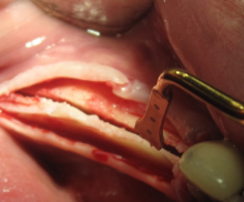
Ridge splitting

  • Ridge splitting with cerabone® - Vafa Moshirabadi, Iran
    Ridge splitting with a piezo instrument
For a stable placement of the implants and a successful general outcome, the alveolar bone width should be larger than 6 mm; this ensures that the implants are covered by at least 1–1.5 mm of bone on both the buccal and palatal sides. Because tooth extraction often leads to a severe bone reduction, the ideal width is not always maintained. When dealing with a severe horizontal deficiency, the application of the GBR technique may thus be limited and challenging, this possibly causing a delay in the implantation. Block grafting is a valid alternative, but is time consuming and may be associated with a higher patient morbidity when autogenous blocks are employed. To address these issues, clinicians have developed a variety of techniques for alveolar ridge splitting. These include the longitudinal splitting of the ridge that makes use of microsaws, diamond discs, or piezosurgical devices to separate the buccal from the palatal plate; a chisel is typically employed to spread them apart. The space created between the plates is then filled with a bone grafting material.
botiss maxresorb®, Jason fleece® and collprotect® membrane for sinus lift and simultaneous implantation - case by Dr. F. Kistler

DVT image demonstrating horizontal and vertical amount of bone available

botiss cerabone for ridge splitting and augmentation of an atrophic alveolar ridge - Clinical case by Dr. V. Moshirabadi

Situation before augmentation, atrophic alveolar ridge

Lateral application of grafting material

The lateral application of a bone substitute material can help stabilize the bone splitting, i.e., the bone plates. In this case, the prior application of a dry membrane is recommended to facilitate the insertion of the particulate grafting material.

Fracture of vestibular plate

A fracture of the vestibular plate is the most common complication in the course of a ridge splitting. Following the implantation, the fractured lamella can be fixed with an osteosynthesis screw.

Ridge splitting with a microwsaw - PD Dr. J. Neugebauer
Kieferkammspaltung mit einer Mikrosäge - PD Dr. J. Neugebauer

The application of a bone substitute material like maxresorb® can help prevent the formation of soft tissue in between the cortical plates. Compared to bovine bone, the biphasic synthetic maxresorb® granules offer the advantage of a complete resorption as they are replaced by the patient's own bone in 2–3 years. In addition, a lateral application of the grafting material can improve the stabilization of the bone splitting.

Furthermore, the use of a membrane, such as the Jason® membrane or collprotect® membrane, to cover the augmentation site is recommended to ensure an undisturbed osseous regeneration. Particularly in the case of a thin biotype, the low thickness of the Jason® membrane facilitates the tension-free closure of the flap over the grafting site.

The expansion of the ridge requires a sufficient flexibility of the bone lamellae. Therefore, this technique is only indicated in the case of a moderate reconstructive need (inside contour <8 mm, outside <4 mm to augment) and minimal ridge width of 2–3 mm; this ensures the presence of cancellous bone between the lamellar plates. The suitable time point of implantation (e.g., whether performed in a one-stage procedure or following a proper amount of healing time) depends on the stability of the expanded segment and the residual bone volume.


Please Contact us for Literature.