Peri-implant soft tissue thickening with mucoderm® - Dr. F. Rojas-Vizcaya
-
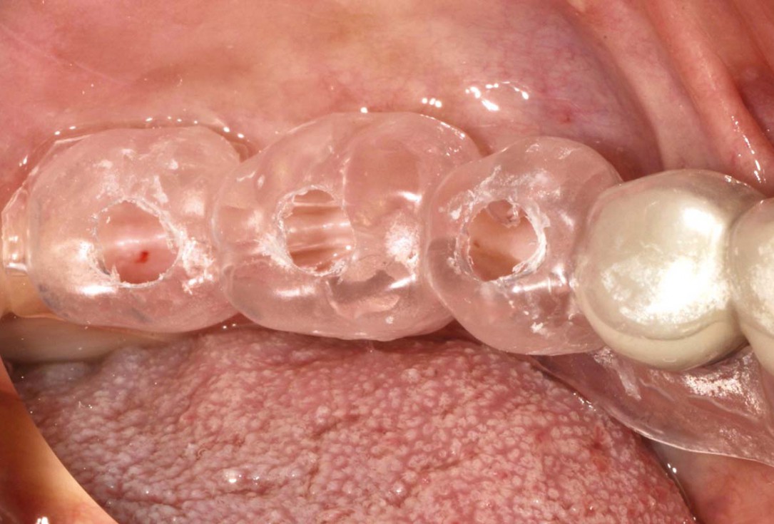
1/16 - Drilling template for guided implant placementPeri-implant soft tissue thickening with mucoderm® - Dr. F. Rojas-Vizcaya
-
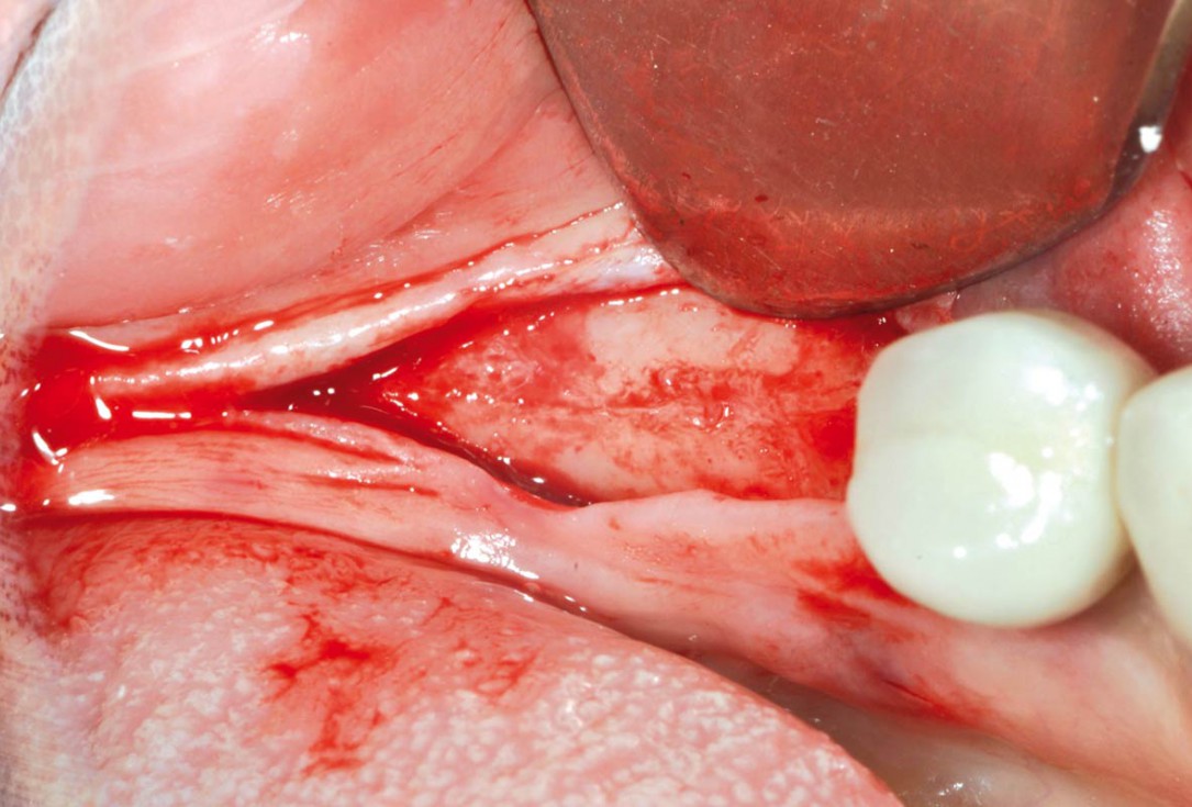
2/16 - Crestal incision and preparation of a mucosal flapPeri-implant soft tissue thickening with mucoderm® - Dr. F. Rojas-Vizcaya
-
3/16 - Implant bed preparationPeri-implant soft tissue thickening with mucoderm® - Dr. F. Rojas-Vizcaya
-
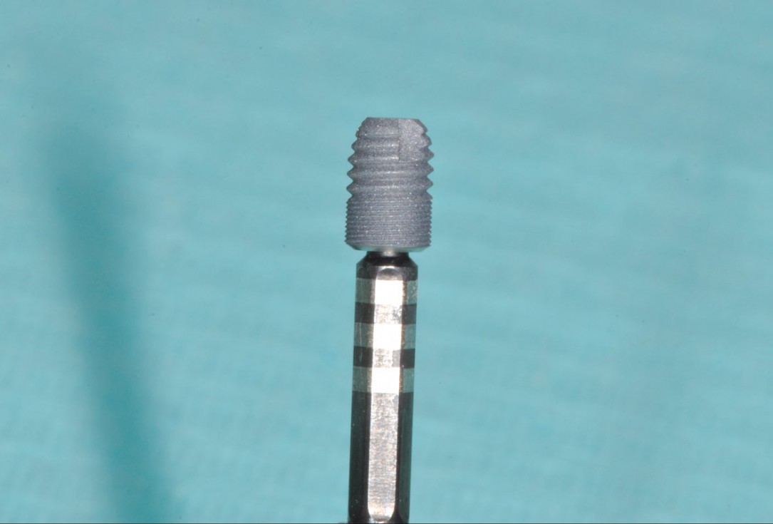
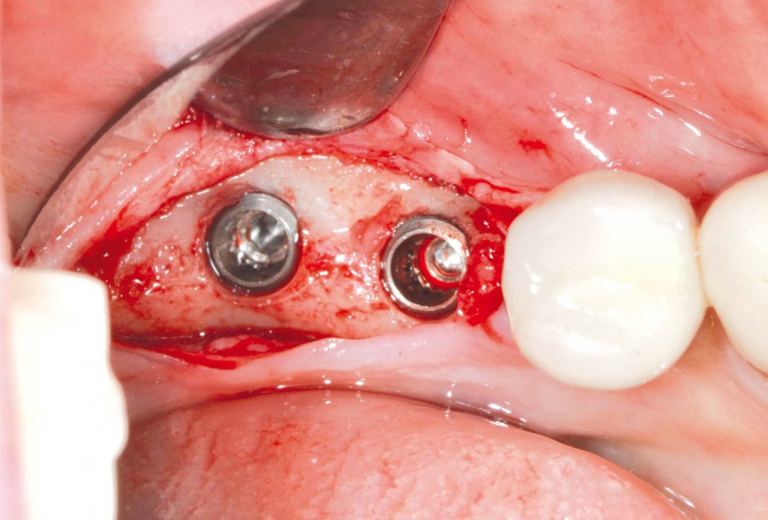
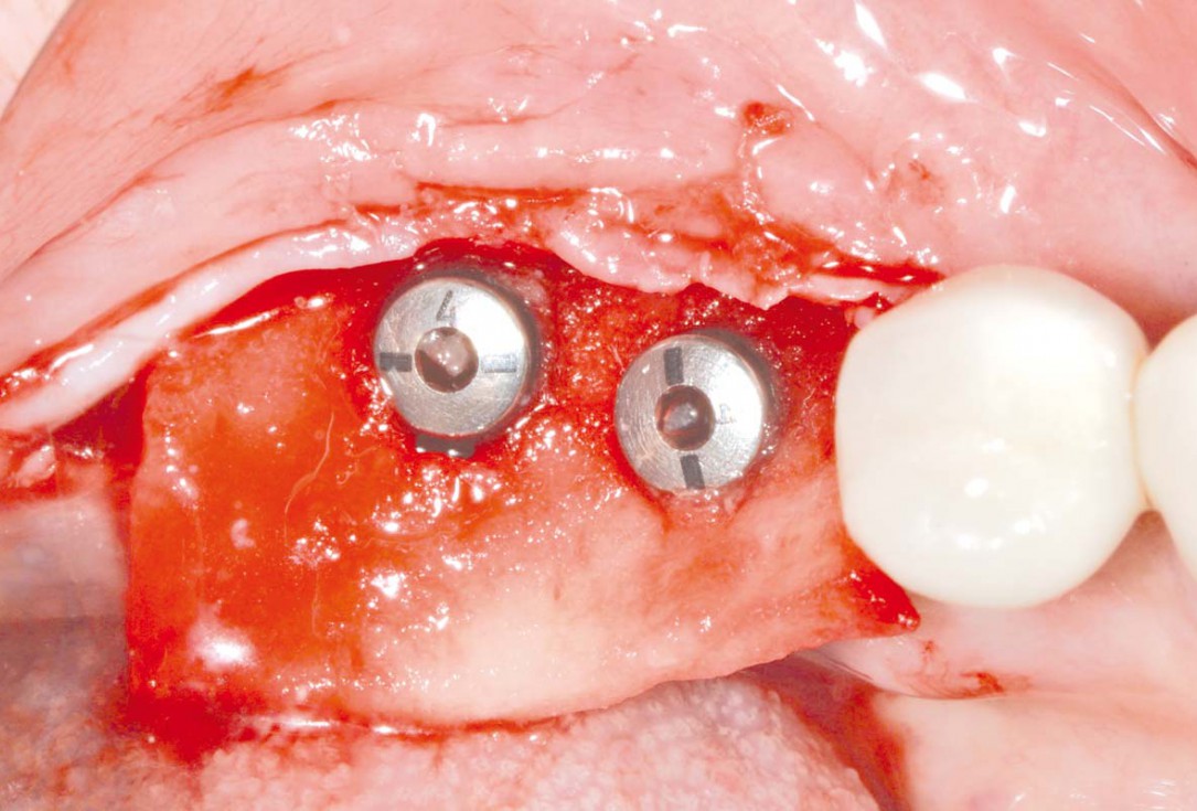
4/16 - Placement of short implantsPeri-implant soft tissue thickening with mucoderm® - Dr. F. Rojas-Vizcaya
-
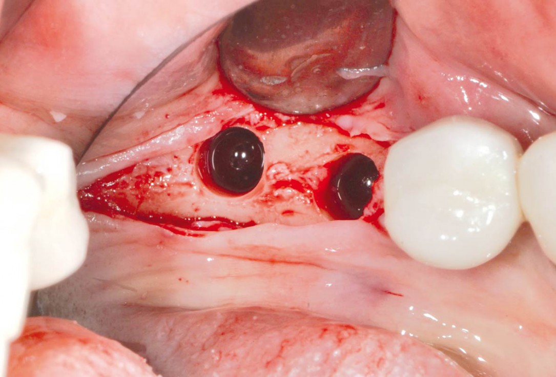
5/16 - Placed implantsPeri-implant soft tissue thickening with mucoderm® - Dr. F. Rojas-Vizcaya
-
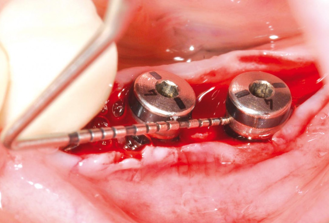
6/16 - Measurement of implant distancePeri-implant soft tissue thickening with mucoderm® - Dr. F. Rojas-Vizcaya
-
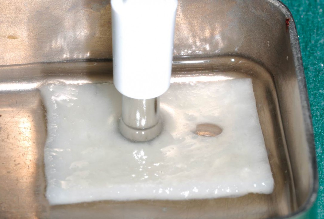
7/16 - Punching of the hydrated mucoderm®Peri-implant soft tissue thickening with mucoderm® - Dr. F. Rojas-Vizcaya
-
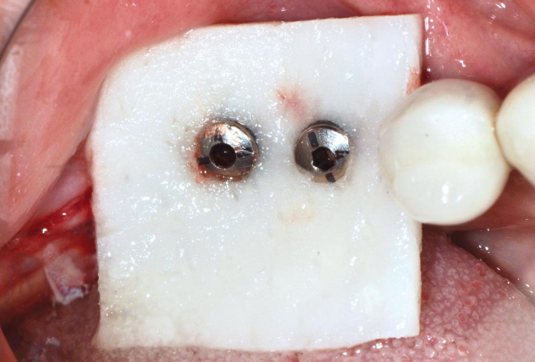
8/16 - mucoderm® pulled over the healing capsPeri-implant soft tissue thickening with mucoderm® - Dr. F. Rojas-Vizcaya
-
9/16 - Adaptation of mucoderm®Peri-implant soft tissue thickening with mucoderm® - Dr. F. Rojas-Vizcaya
-
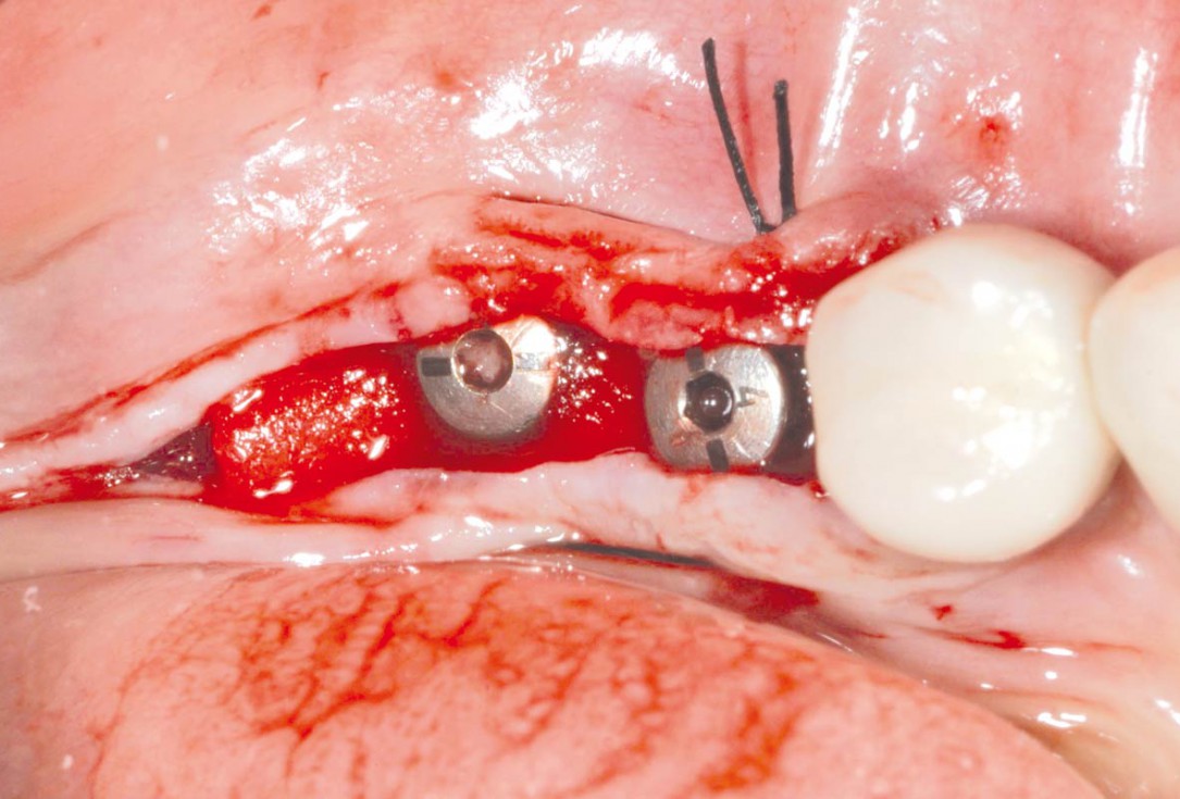
10/16 - Fixation of mucoderm®Peri-implant soft tissue thickening with mucoderm® - Dr. F. Rojas-Vizcaya
-
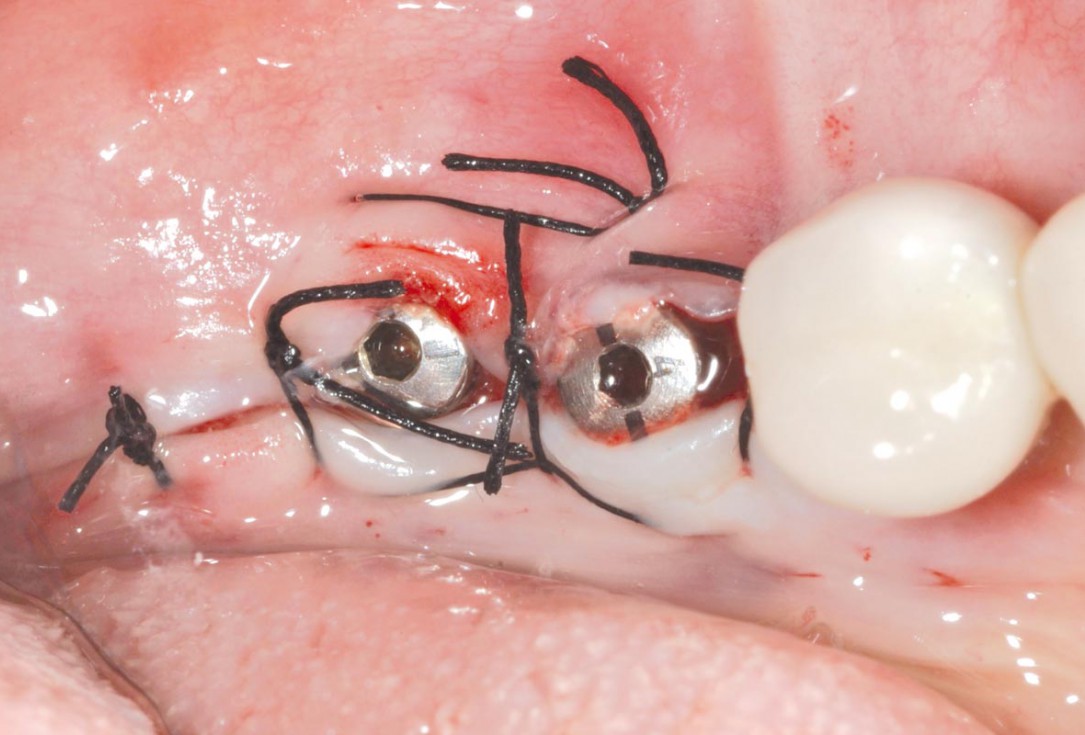
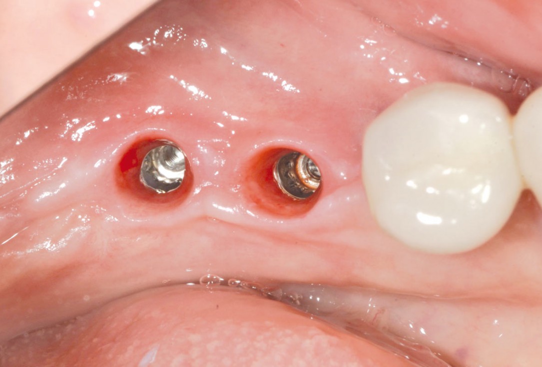
11/16 - Wound closurePeri-implant soft tissue thickening with mucoderm® - Dr. F. Rojas-Vizcaya
-
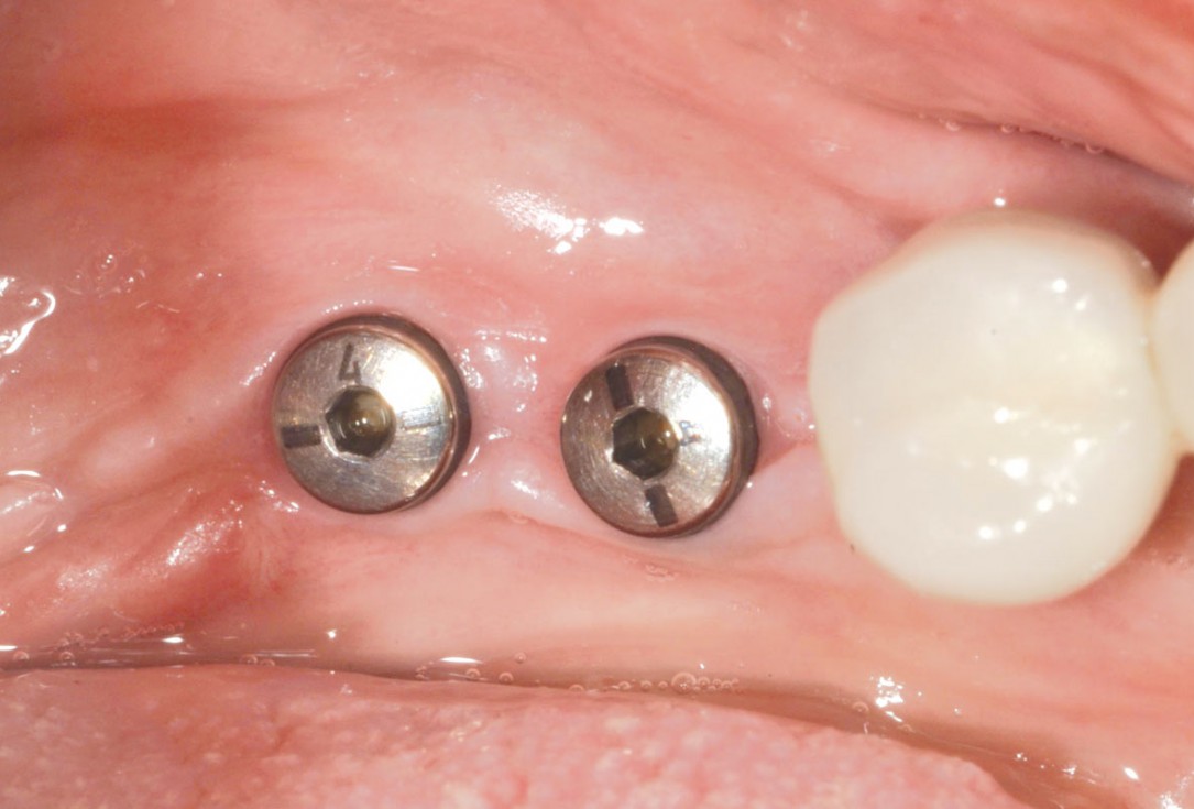
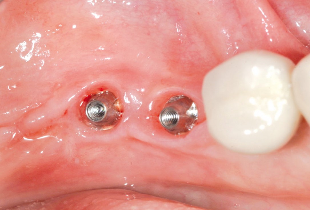
12/16 - Healing at 7 weeksPeri-implant soft tissue thickening with mucoderm® - Dr. F. Rojas-Vizcaya
-
13/16 - Soft tissue profile at 7 weeksPeri-implant soft tissue thickening with mucoderm® - Dr. F. Rojas-Vizcaya
-
14/16 - Nice soft tissue profile for final restorationPeri-implant soft tissue thickening with mucoderm® - Dr. F. Rojas-Vizcaya
-
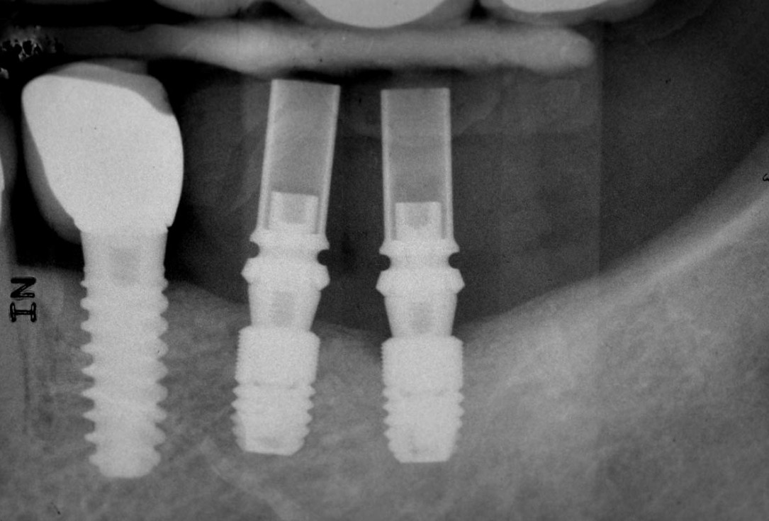
15/16 - Post-surgical x-rayPeri-implant soft tissue thickening with mucoderm® - Dr. F. Rojas-Vizcaya
-
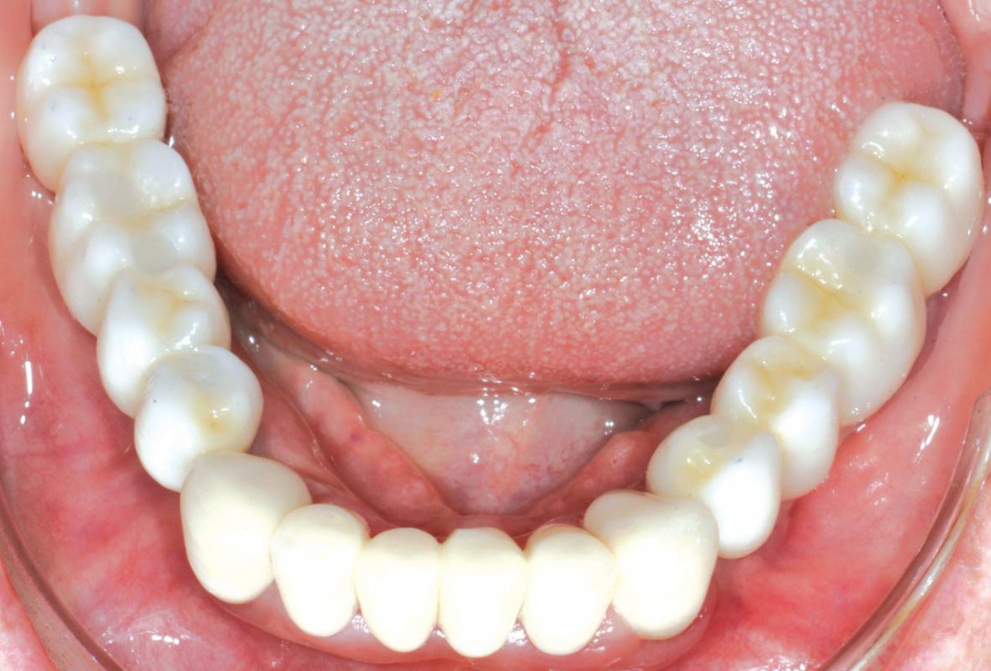
16/16 - Placement of definitive crownsPeri-implant soft tissue thickening with mucoderm® - Dr. F. Rojas-Vizcaya

Initial view of the case. Discoloration of 1.1 and mild class I gingival recession

Initial clinical situation with Miller class 1 recession

Initial clinical situation

Initial clinical situation showing strongly compromised tooth 21

Pre-surgical clinical situation. Deep gingival recessions at both upper canine.

Pre-surgical situation. Multiple adjacent gingival recessions at teeth 12, 13 and 14.

Multiple adjacent gingival recessions.

X-ray shows a 3-dimensional periondontal defect

Initial clinical situation with lack of keratinized tissue

Bone defect in area 11-21 due to two lost implants (periimplantitis) after 15 years of function

Intact socket following atraumatic tooth extraction

Full-thickness flap preparation bucally and lingually

Initial situation: missing teeth #11 & 12 and badly broken #21 root

Alveolar socket before soft and hard tissue augmentation

Clinical situation before surgery

Initial clinical situation

Initial clinical situation with traumatic loss of tooth 21

Longitudinal fracture on the root resected tooth 21 with visible buccal fistula

X-ray showing endodontic failure of the molar

X-ray of initial clinical situation

Initial clinical situation

Initial clinical situation

Pre-operative clinical situation. Gingival recessions at teeth 11 and 21.

Gingival recession at tooth 13. Free gingival graft (FGG) of a previous surgery for root coverage visible.

recession on tooth 11

Insufficient keratinized mucosa and extremely shallow vestibule on the maxilla

Initial clinical situation

Tooth extraction due to root fracture
