Mucosal thickening around bone level implants - Dr. A. Puisys
-
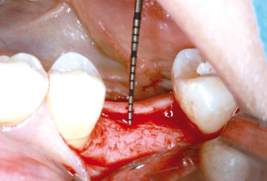
1/10 - Full-thickness flap preparation bucally and linguallyMucosal thickening around bone level implants - Dr. A. Puisys
-
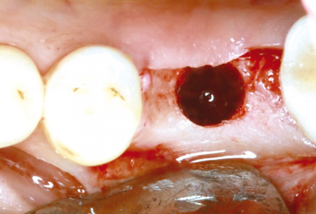
2/10 - Bone preparation for Straumann® bone level implantMucosal thickening around bone level implants - Dr. A. Puisys
-
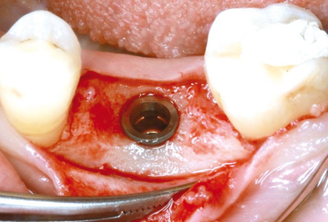
3/10 - Implant insertion and contouring crestal bone with straight handpieceMucosal thickening around bone level implants - Dr. A. Puisys
-
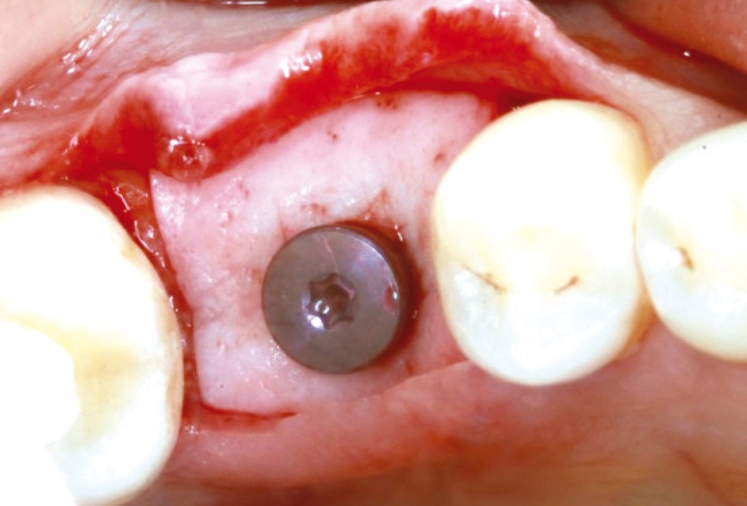
4/10 - Rehydrated mucoderm® perforated and pulled over the healing capMucosal thickening around bone level implants - Dr. A. Puisys
-
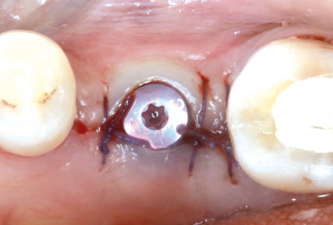
5/10 - The margins of the flap are adapted and sutured leaving the abutment openMucosal thickening around bone level implants - Dr. A. Puisys
-
6/10 - Situation 1 week postoperative and after suture removalMucosal thickening around bone level implants - Dr. A. Puisys
-
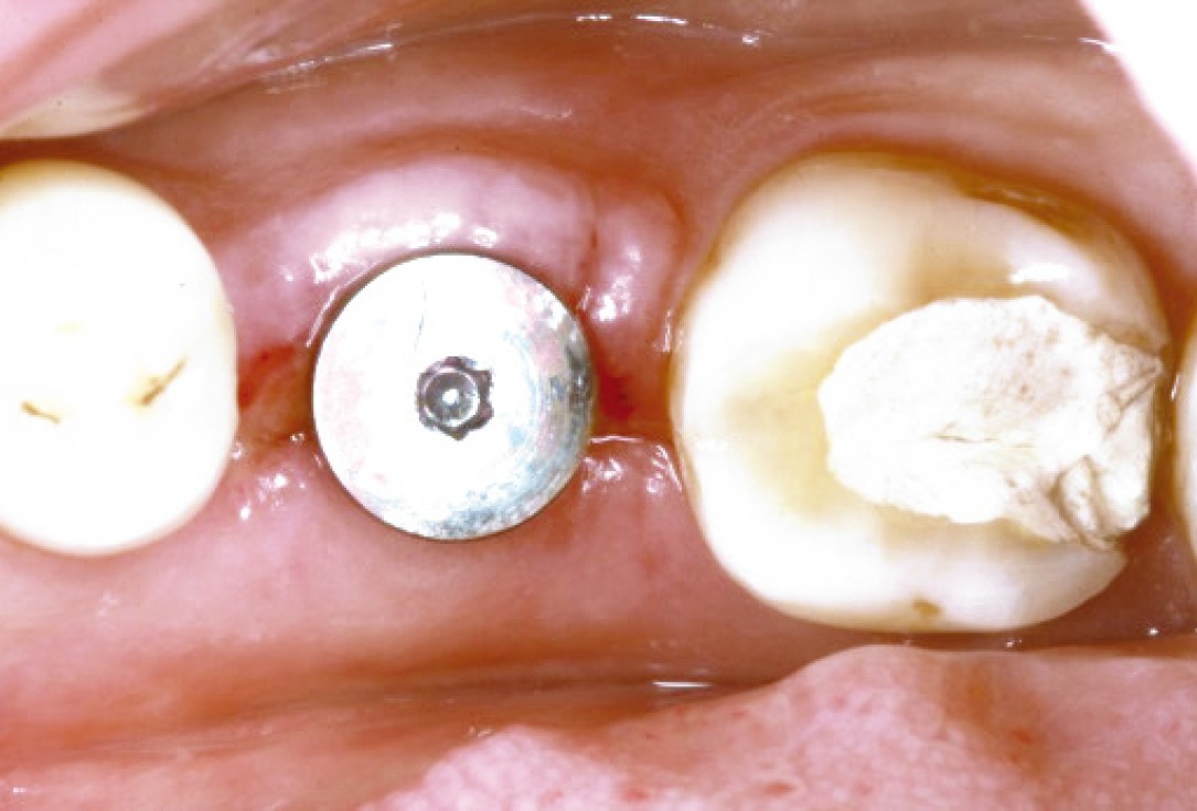
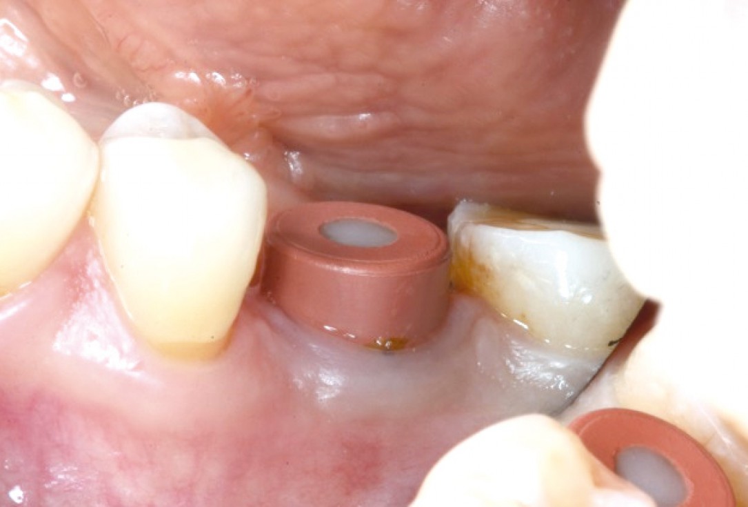
7/10 - Wider healing abutment after 4 monthsMucosal thickening around bone level implants - Dr. A. Puisys
-
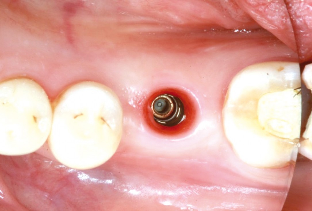
8/10 - Smooth emergence profile visible after removal of the healing abutmentMucosal thickening around bone level implants - Dr. A. Puisys
-
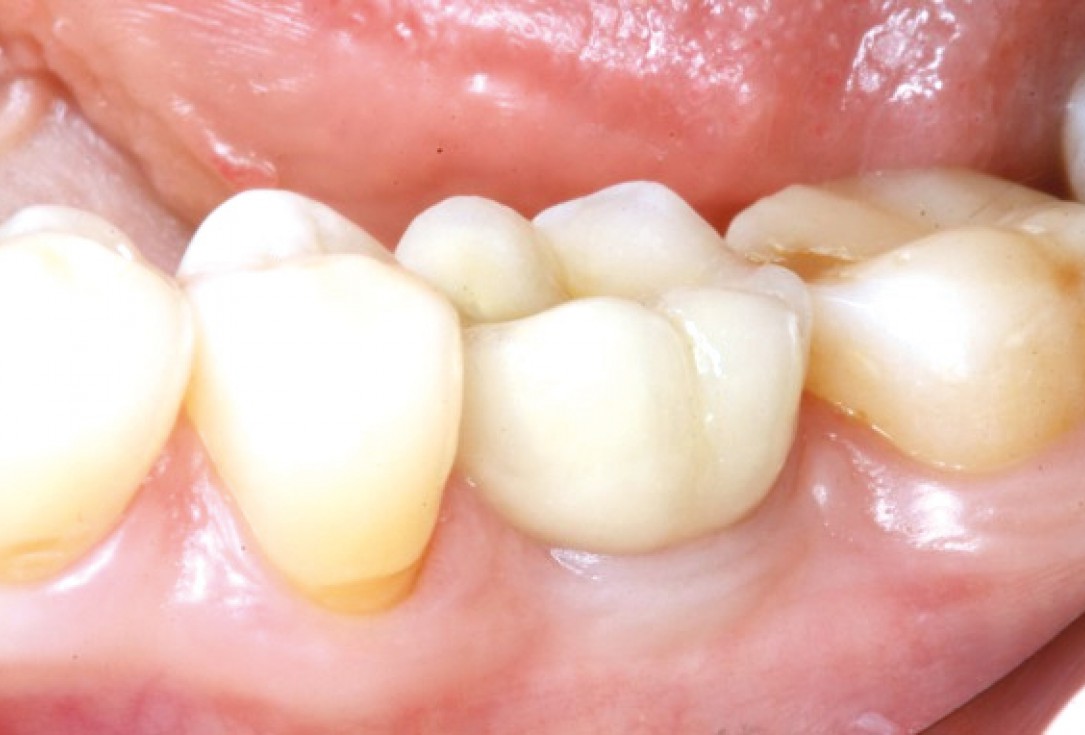
9/10 - Final restoration 5 months post-operativeMucosal thickening around bone level implants - Dr. A. Puisys
-
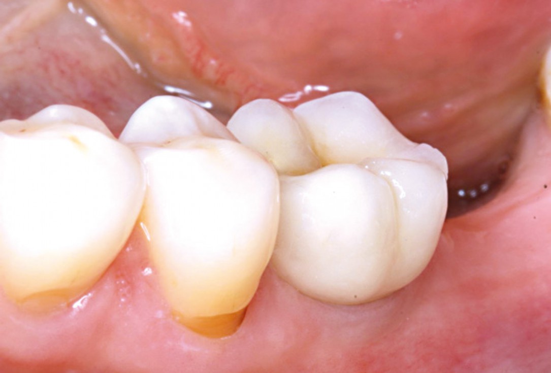
10/10 - Stable clinical situation after 5 yearsMucosal thickening around bone level implants - Dr. A. Puisys

Initial clinical situation

Initial clinical situation

Initial clinical situation

Drilling template for guided implant placement

recession on tooth 11

Initial clinical situation

Initial clinical situation with narrow ridge

Initial clinical situation showing tooth 45 not worth preserving

X-ray of initial clinical situation

Initial clinical situation showing severe soft tissue loss

Initial clinical situation

X-ray shows a 3-dimensional periondontal defect

Bone defect in area 11-21 due to two lost implants (periimplantitis) after 15 years of function

Probing demonstrates peri-implant pocket depth of 8 mm
