Peri-implant soft tissue thickening with mucoderm® - Dr. F. Rojas-Vizcaya
-
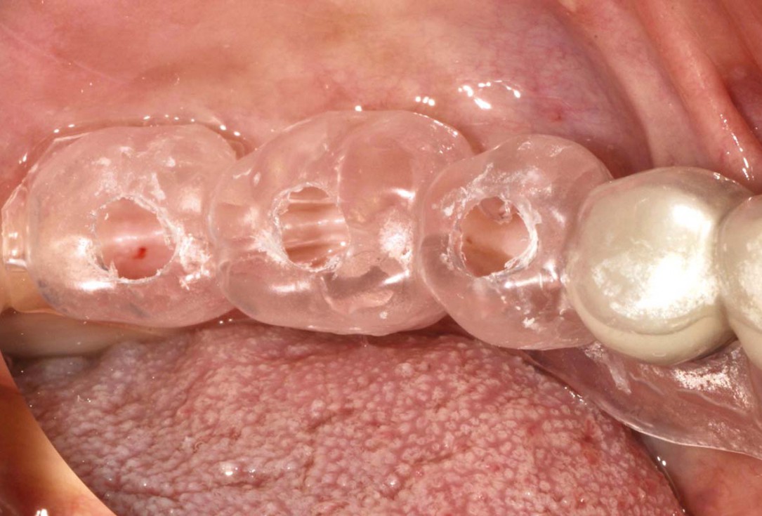
1/16 - Drilling template for guided implant placementPeri-implant soft tissue thickening with mucoderm® - Dr. F. Rojas-Vizcaya
-
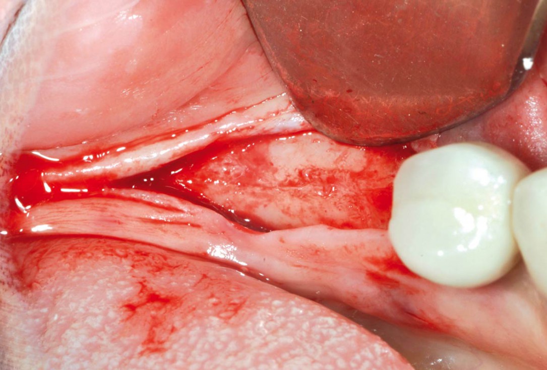
2/16 - Crestal incision and preparation of a mucosal flapPeri-implant soft tissue thickening with mucoderm® - Dr. F. Rojas-Vizcaya
-
3/16 - Implant bed preparationPeri-implant soft tissue thickening with mucoderm® - Dr. F. Rojas-Vizcaya
-
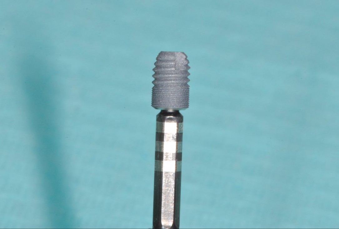
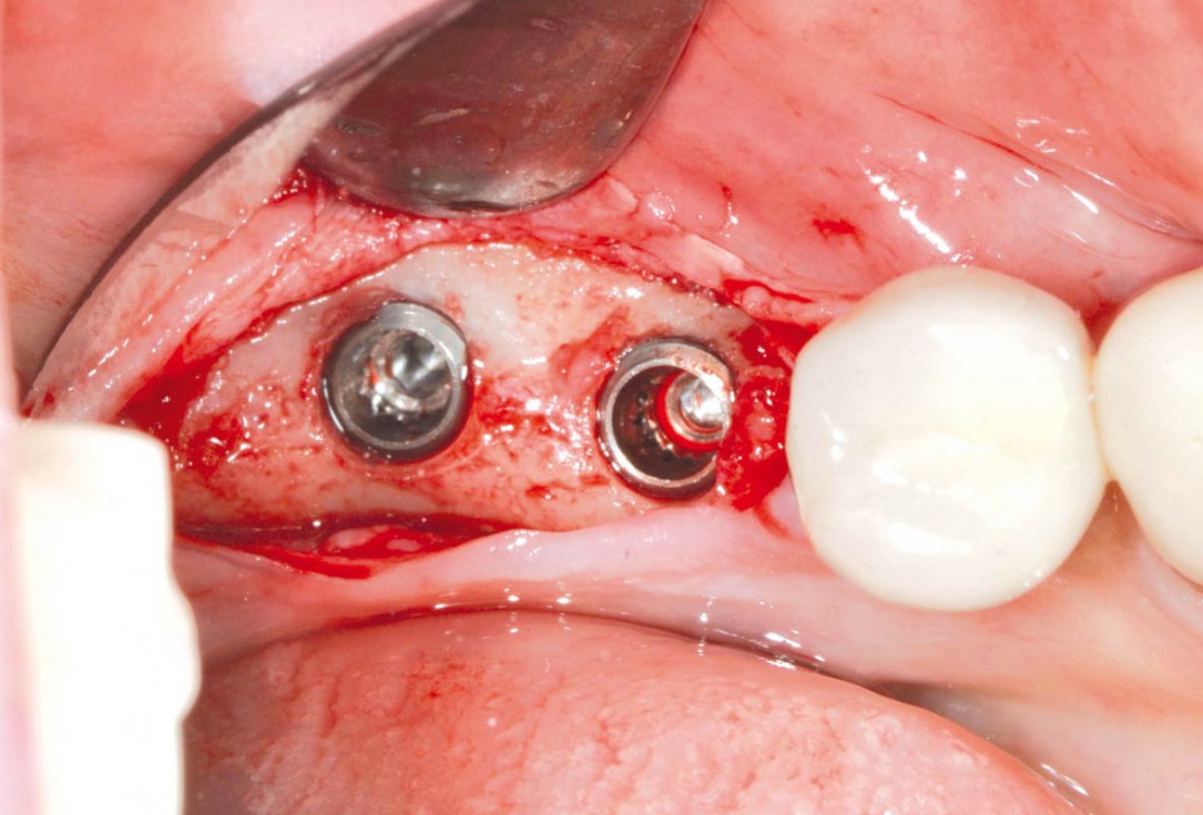
4/16 - Placement of short implantsPeri-implant soft tissue thickening with mucoderm® - Dr. F. Rojas-Vizcaya
-
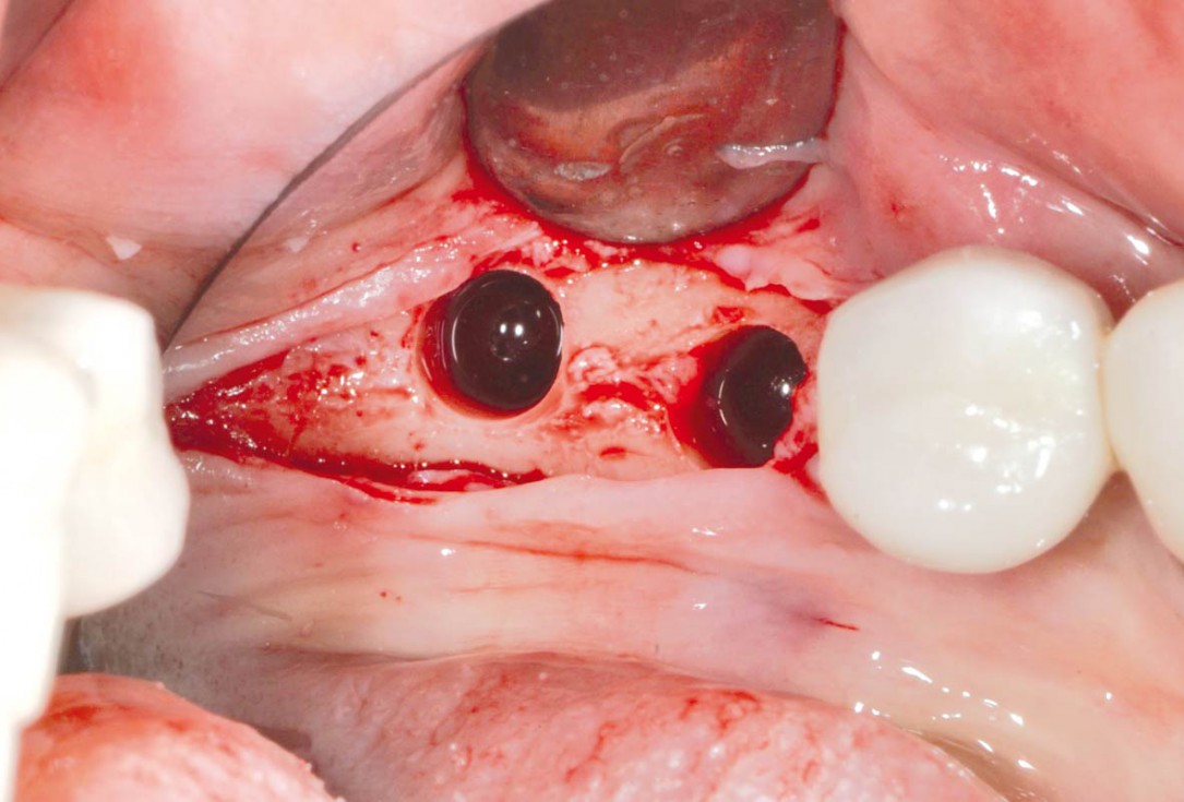
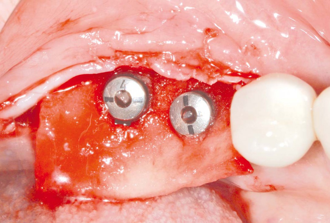
5/16 - Placed implantsPeri-implant soft tissue thickening with mucoderm® - Dr. F. Rojas-Vizcaya
-
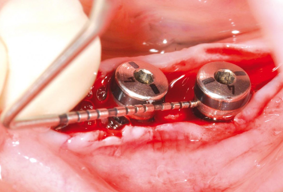
6/16 - Measurement of implant distancePeri-implant soft tissue thickening with mucoderm® - Dr. F. Rojas-Vizcaya
-
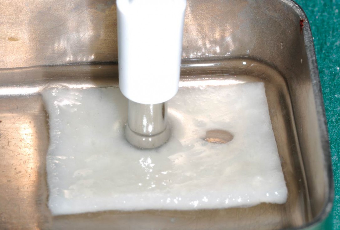
7/16 - Punching of the hydrated mucoderm®Peri-implant soft tissue thickening with mucoderm® - Dr. F. Rojas-Vizcaya
-
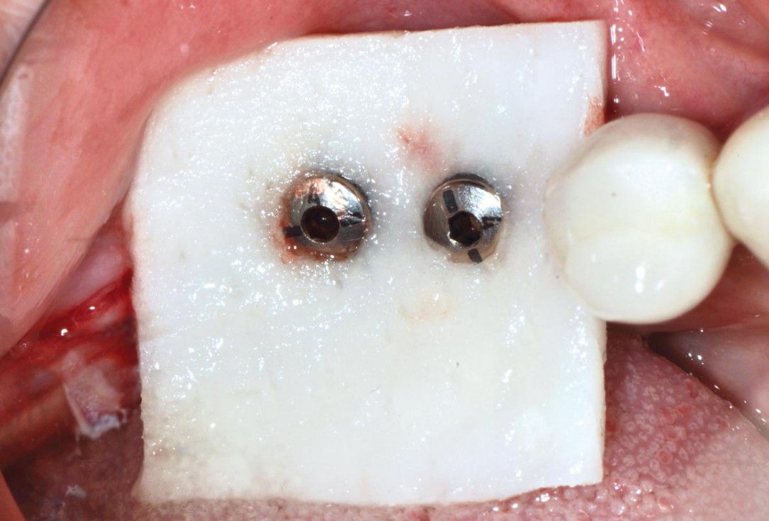
8/16 - mucoderm® pulled over the healing capsPeri-implant soft tissue thickening with mucoderm® - Dr. F. Rojas-Vizcaya
-
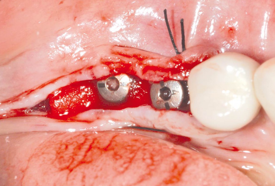
9/16 - Adaptation of mucoderm®Peri-implant soft tissue thickening with mucoderm® - Dr. F. Rojas-Vizcaya
-
10/16 - Fixation of mucoderm®Peri-implant soft tissue thickening with mucoderm® - Dr. F. Rojas-Vizcaya
-
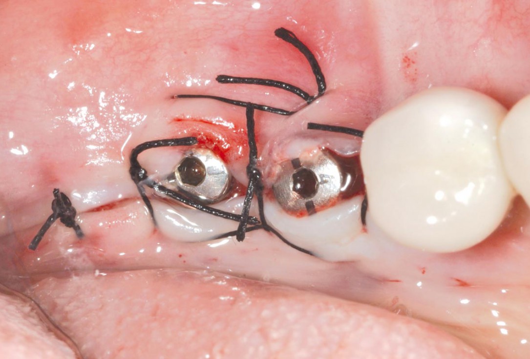
11/16 - Wound closurePeri-implant soft tissue thickening with mucoderm® - Dr. F. Rojas-Vizcaya
-
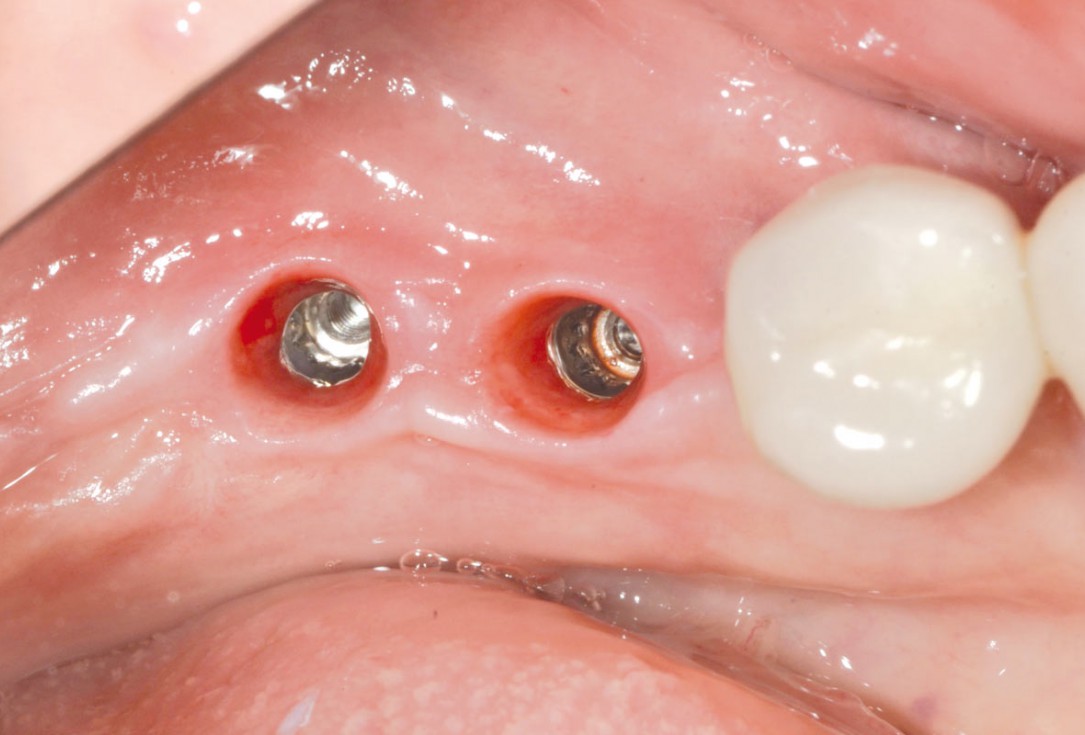
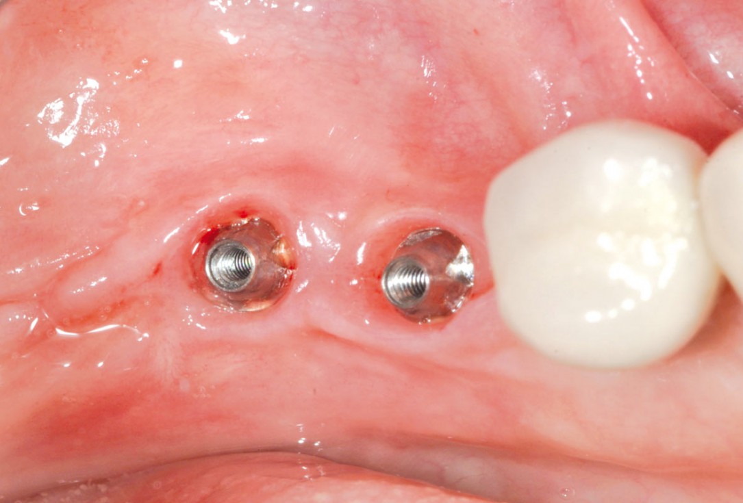
12/16 - Healing at 7 weeksPeri-implant soft tissue thickening with mucoderm® - Dr. F. Rojas-Vizcaya
-
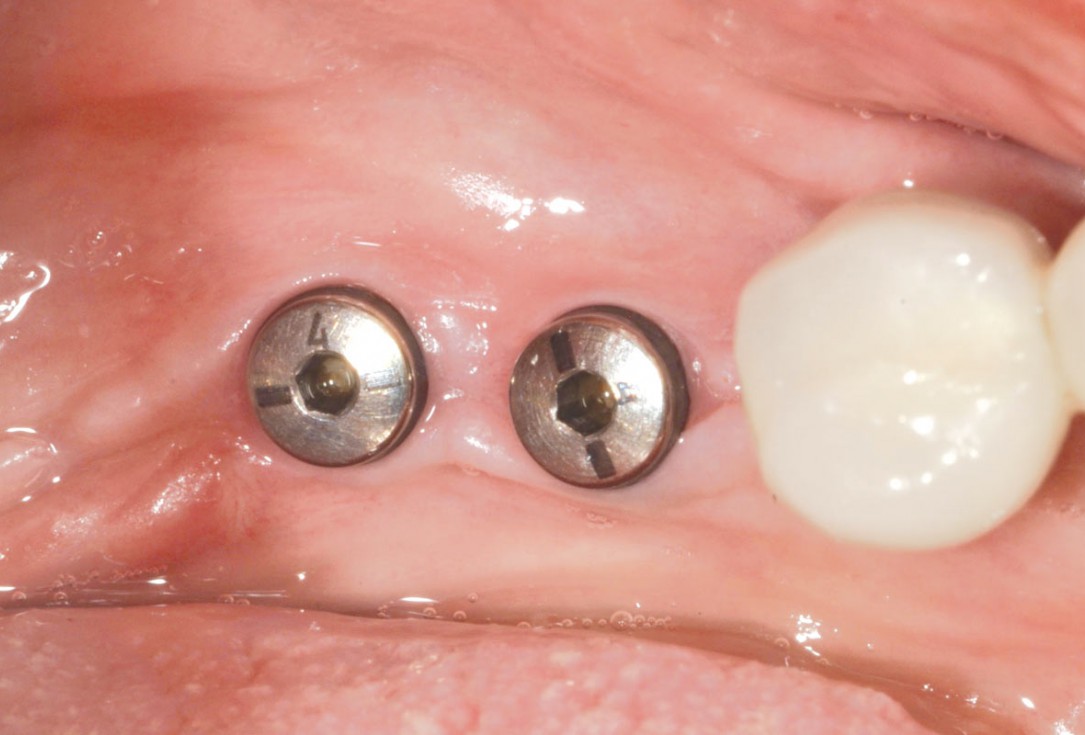
13/16 - Soft tissue profile at 7 weeksPeri-implant soft tissue thickening with mucoderm® - Dr. F. Rojas-Vizcaya
-
14/16 - Nice soft tissue profile for final restorationPeri-implant soft tissue thickening with mucoderm® - Dr. F. Rojas-Vizcaya
-
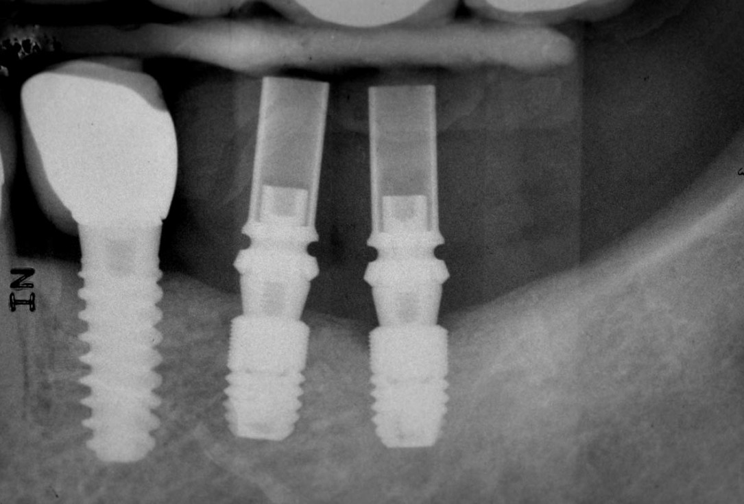
15/16 - Post-surgical x-rayPeri-implant soft tissue thickening with mucoderm® - Dr. F. Rojas-Vizcaya
-
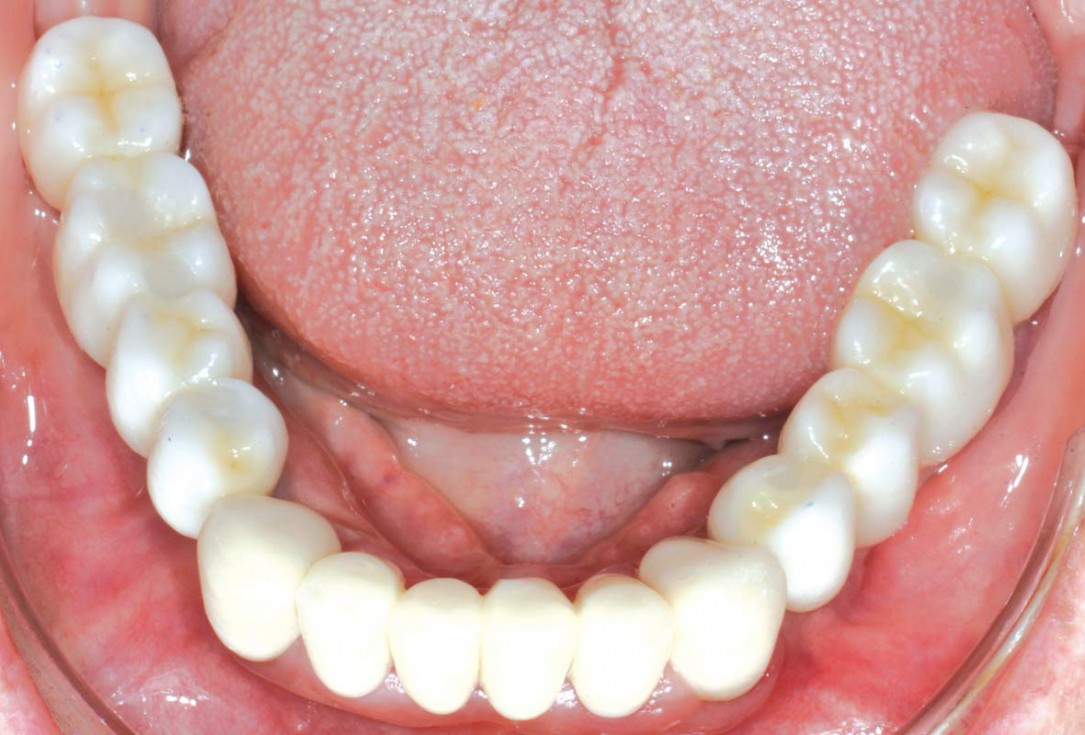
16/16 - Placement of definitive crownsPeri-implant soft tissue thickening with mucoderm® - Dr. F. Rojas-Vizcaya

Initial clinical situation

Initial clinical situation

Initial clinical situation

Full-thickness flap preparation bucally and lingually

recession on tooth 11

Initial clinical situation

Initial clinical situation with narrow ridge

Initial clinical situation showing tooth 45 not worth preserving

X-ray of initial clinical situation

Initial clinical situation showing severe soft tissue loss

Initial clinical situation

X-ray shows a 3-dimensional periondontal defect

Bone defect in area 11-21 due to two lost implants (periimplantitis) after 15 years of function

Probing demonstrates peri-implant pocket depth of 8 mm
