Immediate implantation with maxresorb® - Dr. M. Frosecchi
-
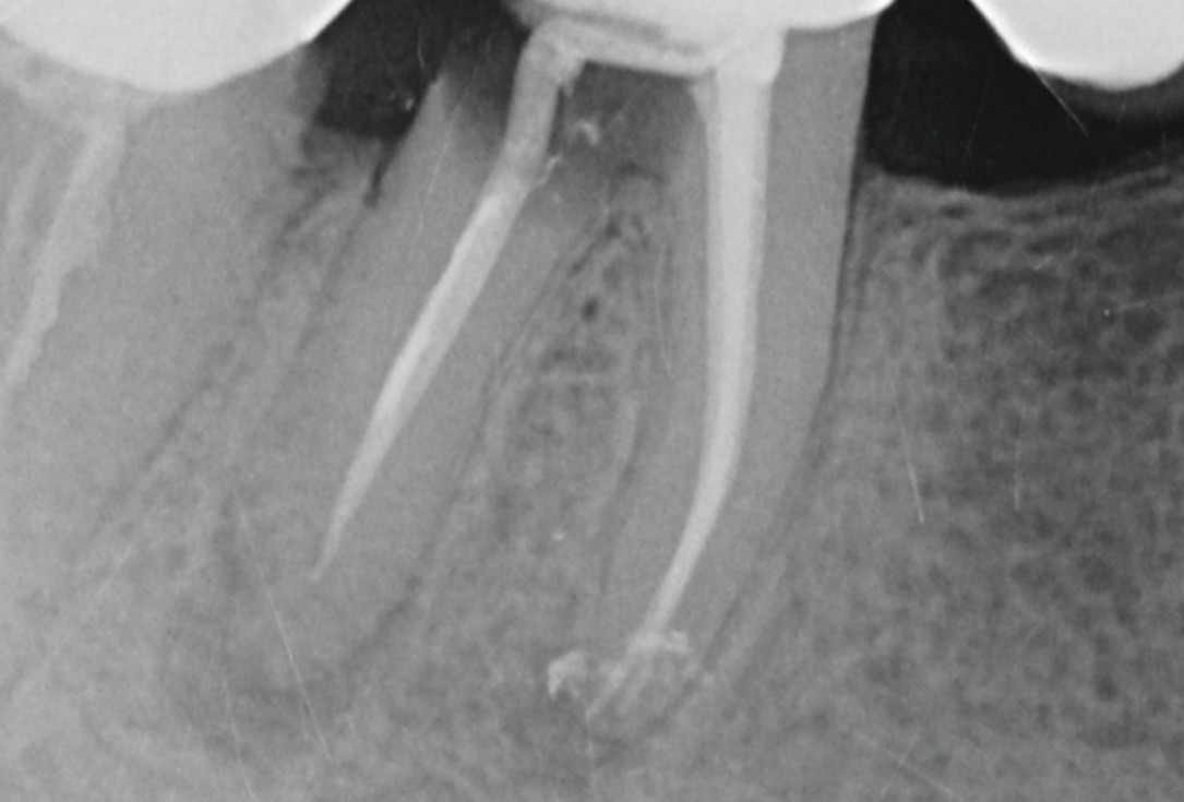
01/11 - X-ray control before tooth extractionImmediate implantation with maxresorb® - Dr. M. Frosecchi
-
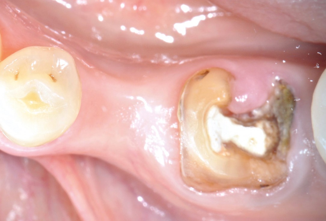
02/11 - Clinical situation before tooth extractionImmediate implantation with maxresorb® - Dr. M. Frosecchi
-
03/11 - Situation after extraction of tooth 35 and flap preparationImmediate implantation with maxresorb® - Dr. M. Frosecchi
-
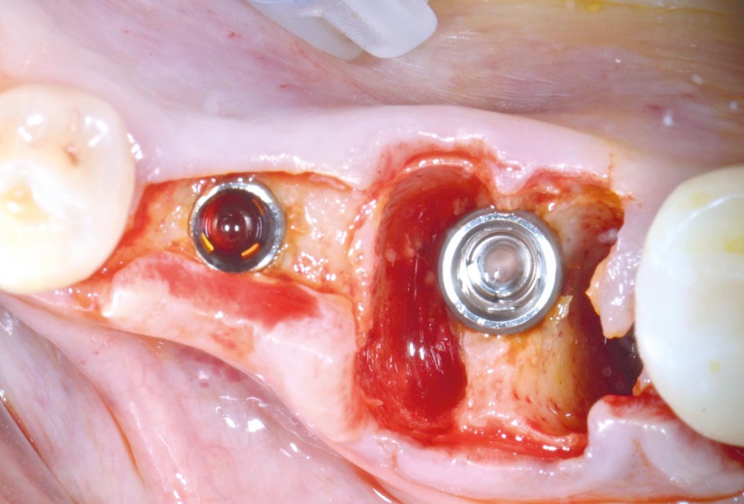
04/11 - Implant insertion in position 35 and 34Immediate implantation with maxresorb® - Dr. M. Frosecchi
-
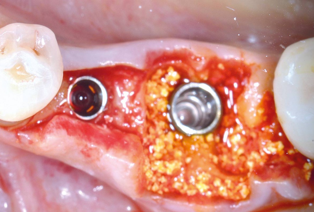
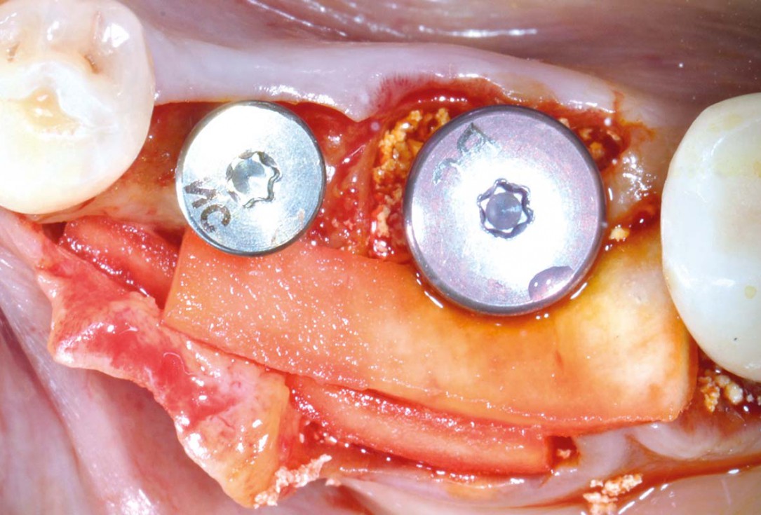
05/11 - Filling of gaps around immediately placed implant with maxresorb®Immediate implantation with maxresorb® - Dr. M. Frosecchi
-
06/11 - Positioning of mucoderm® at buccal side for soft tissue thickeningImmediate implantation with maxresorb® - Dr. M. Frosecchi
-
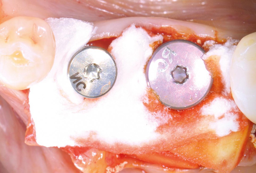
07/11 - Covering with a collagen fleeceImmediate implantation with maxresorb® - Dr. M. Frosecchi
-
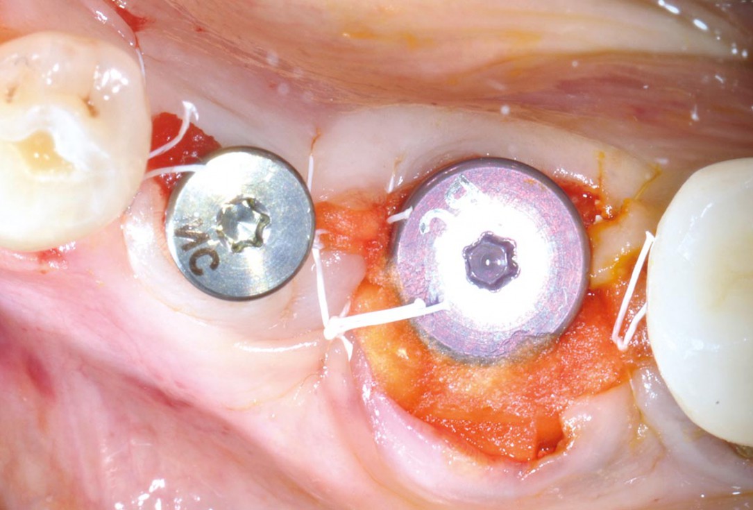
08/11 - Flap adaptation and suturing, fleece partially left exposedImmediate implantation with maxresorb® - Dr. M. Frosecchi
-
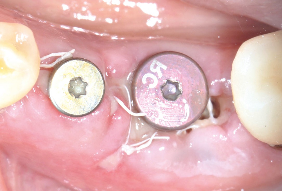
09/11 - Clinical situation 7 days post-opImmediate implantation with maxresorb® - Dr. M. Frosecchi
-
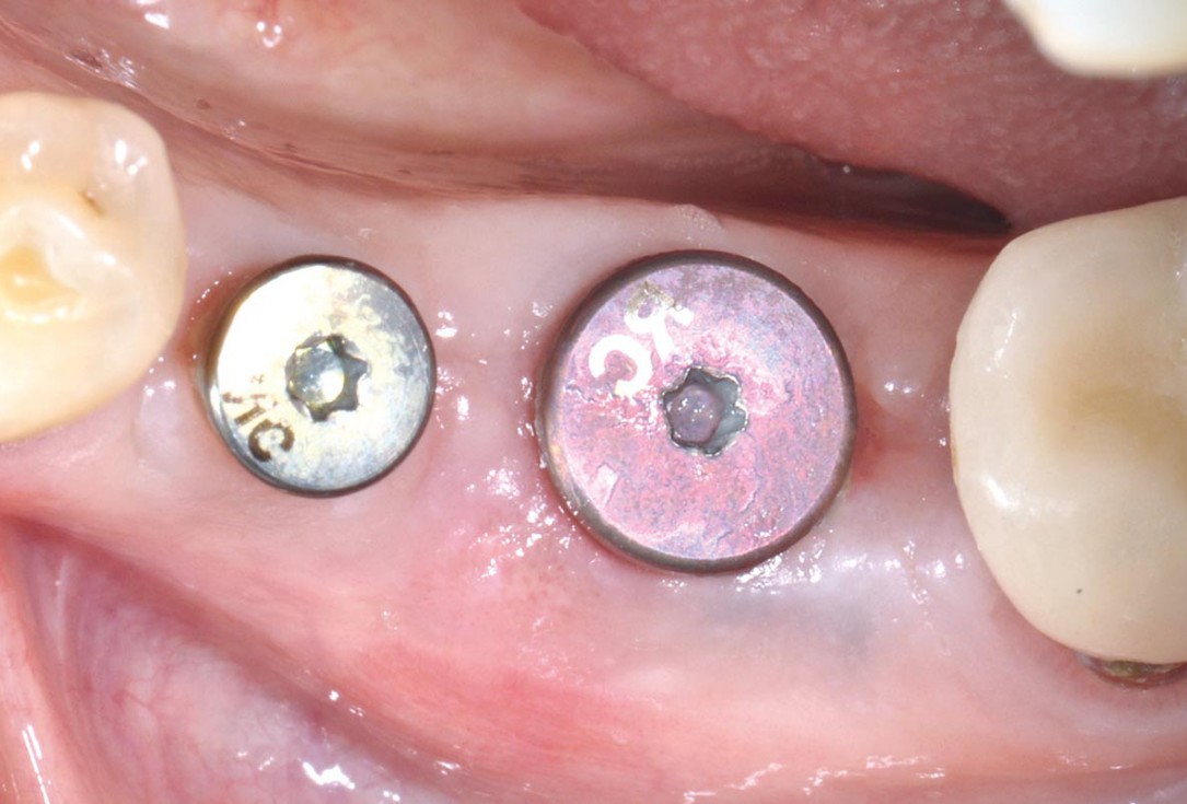
10/11 - Healing 3 months post-opImmediate implantation with maxresorb® - Dr. M. Frosecchi
-
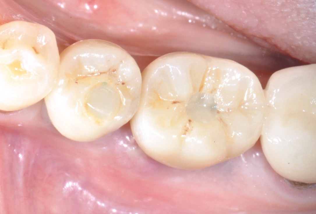
11/11 - Final restoration with screw-retained zirkonium crownsImmediate implantation with maxresorb® - Dr. M. Frosecchi

Initial Orthopantomograph X-Ray

DVT image showing the reduced amount of bone available in the area of the mental foramen

X-ray shows a 3-dimensional periondontal defect

Initial situation: Inflammated tooth #12

Clinical situation before extraction and implantation

Pre-operative OPG, tooth 25 planned for extraction

Full-thickness flap preparation bucally and lingually

Initial situation: missing teeth #11 & 12 and badly broken #21 root

Drilling template for guided implant placement

Initial clinical situation showing strongly compromised tooth 21

Surgical presentation of the alveolar ridge with reduced amount of horizontal bone available

Pre-operative x-ray

Clinical situation before extraction

DVT image demonstrating horizontal and vertical amount of bone available

Preoperative clinical situation

DVT control after sinusitis surgery, residual bone height 1 mm

Preoperative CBCT analysis

X-ray of initial clinical situation

Initial clinical situation

Alveolar socket before soft and hard tissue augmentation

Surgical presentation of the alveolar ridge with reduced amount of horizontal bone available

DVT control after sinusitis surgery, residual bone height 1 mm

X-ray control showing initial situation

DVT control after sinusitis surgery, residual bone height 1 mm

Pre-operative situation showing tooth 21 with deep periodontal pocket. Tooth presented with mobility grade III.

Analysis of the situation and planning of the implant positions based on CBCT

Initial view of the case. Discoloration of 1.1 and mild class I gingival recession

Initial clinical situation with Miller class 1 recession

Initial clinical situation

Initial clinical situation
